
Fig. S15
- ID
- ZDB-FIG-200306-137
- Publication
- Nimura et al., 2019 - Role of Reelin in cell positioning in the cerebellum and the cerebellum-like structure in zebrafish
- Other Figures
- All Figure Page
- Back to All Figure Page
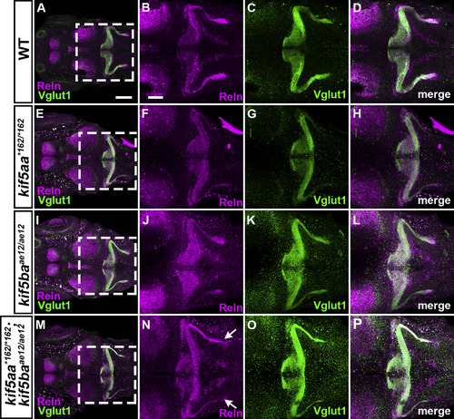
S15. Reln and GC axons in kinesin mutants. WT (A-D, n= 4), kif5aa*162/*162 (E-H, n= 13), kif5baae12/ae12 (I-L, n= 4), and kif5aa*162/*162; kif5baae12/ae12 (M-P, n= 7) larvae at 5-dpf were stained with anti-Reln (magenta) and anti-Vglut1 (green) antibodies. Dorsal views with anterior to the left. (B-D, F–H, J-L, N–P) High magnification images of the boxes in A, E, I, and M. Typical images are shown. Note that the Reln and Vglut1 stainings were not strongly affected in the kif5aa*162/*162 and kif5baae12/ae12single mutants, whereas they were decreased in the double mutants (indicated by arrows in N). Detailed analysis of the double mutants is shown in Fig. S1. Scale bars: 200 μm in A (applies to A, E, I, M); 100 μm in B (applies to B-D, F–H, J-L, N–P). |
Reprinted from Developmental Biology, 455(2), Nimura, T., Itoh, T., Hagio, H., Hayashi, T., Di Donato, V., Takeuchi, M., Itoh, T., Inoguchi, F., Sato, Y., Yamamoto, N., Katsuyama, Y., Del Bene, F., Shimizu, T., Hibi, M., Role of Reelin in cell positioning in the cerebellum and the cerebellum-like structure in zebrafish, 393-408, Copyright (2019) with permission from Elsevier. Full text @ Dev. Biol.