
FIGURE
Fig. S9
- ID
- ZDB-FIG-200306-131
- Publication
- Nimura et al., 2019 - Role of Reelin in cell positioning in the cerebellum and the cerebellum-like structure in zebrafish
- Other Figures
- All Figure Page
- Back to All Figure Page
Fig. S9
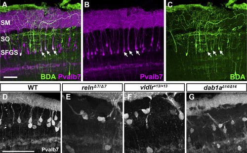
Pvalb7 is a marker for type I neuron. (A-C) Tracing of type I neurons. Biotinylated dextran amine (BDA) was injected into the SM of the tectum. After incubation, transverse sections were labeled with immunofluorescence using anti-Pvalb7 antibody (magenta) and fluorescent streptavidin (green). Note that the neurons labeled with BDA are also positive for Pvalb7 (indicated by arrows). (D-G) High magnification, grayscale images of Pvalb7-positive cells in Fig. 6D, J, P, and V. Scale bars: 50 μm in A (applies to A-C) and D (applies to D-G). |
Expression Data
Expression Detail
Antibody Labeling
Phenotype Data
Phenotype Detail
Acknowledgments
This image is the copyrighted work of the attributed author or publisher, and
ZFIN has permission only to display this image to its users.
Additional permissions should be obtained from the applicable author or publisher of the image.
Reprinted from Developmental Biology, 455(2), Nimura, T., Itoh, T., Hagio, H., Hayashi, T., Di Donato, V., Takeuchi, M., Itoh, T., Inoguchi, F., Sato, Y., Yamamoto, N., Katsuyama, Y., Del Bene, F., Shimizu, T., Hibi, M., Role of Reelin in cell positioning in the cerebellum and the cerebellum-like structure in zebrafish, 393-408, Copyright (2019) with permission from Elsevier. Full text @ Dev. Biol.