
Fig. S8
- ID
- ZDB-FIG-200306-130
- Publication
- Nimura et al., 2019 - Role of Reelin in cell positioning in the cerebellum and the cerebellum-like structure in zebrafish
- Other Figures
- All Figure Page
- Back to All Figure Page
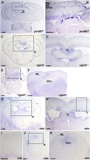
Expression of pvalb7, vglut1, and reln in the mesencephalic tectum and the cerebellum. Cross sections of the mesencephalon (A-D, G-I) and the cerebellum (E, F, J, K) of adult WT zebrafish were stained with antisense riboprobes of pvalb7 (A, B), vglut1 (C–F), or reln (G-K) genes. (B, D, F, H, I, K) High magnification images of the boxes in A, C, E, G, H, and J. Note that pvalb7-expressing cells are located in the SFGS of the tectum (A, B), and reln and vglut1 are not detected in the superficial layer of the tectum and the cerebellum (D, F, H, I). The abbreviations are described in the legend of Fig. 1. Scale bars: 200 μm in A, C, E, G, J; 100 μm in B, D, F, H, I, K. |
Reprinted from Developmental Biology, 455(2), Nimura, T., Itoh, T., Hagio, H., Hayashi, T., Di Donato, V., Takeuchi, M., Itoh, T., Inoguchi, F., Sato, Y., Yamamoto, N., Katsuyama, Y., Del Bene, F., Shimizu, T., Hibi, M., Role of Reelin in cell positioning in the cerebellum and the cerebellum-like structure in zebrafish, 393-408, Copyright (2019) with permission from Elsevier. Full text @ Dev. Biol.