
FIGURE
Fig. S4
- ID
- ZDB-FIG-080715-23
- Publication
- Krens et al., 2008 - Distinct functions for ERK1 and ERK2 in cell migration processes during zebrafish gastrulation
- Other Figures
- All Figure Page
- Back to All Figure Page
Fig. S4
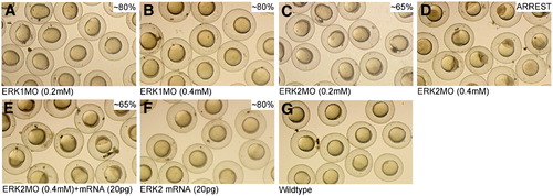
Specific functions of ERK1 and ERK2 revealed by concentration-dependent knockdown during early embryogenesis. Embryos were injected with 0.2 mM (1.7 ng) and 0.4 mM (3.4 ng) ERK1MO (A,B) or ERK2MO (C,D) respectively. Rescue was performed by co-injection of 3.4 ng ERK2MO with 20 pg ERK2 mRNA (E) and compared to phenotypes of 20 pg ERK2 mRNA injected (F) and wild type embryos at 8 hpf (G). |
Expression Data
Expression Detail
Antibody Labeling
Phenotype Data
Phenotype Detail
Acknowledgments
This image is the copyrighted work of the attributed author or publisher, and
ZFIN has permission only to display this image to its users.
Additional permissions should be obtained from the applicable author or publisher of the image.
Reprinted from Developmental Biology, 319(2), Krens, S.F., He, S., Lamers, G.E., Meijer, A.H., Bakkers, J., Schmidt, T., Spaink, H.P., and Snaar-Jagalska, B.E., Distinct functions for ERK1 and ERK2 in cell migration processes during zebrafish gastrulation, 370-383, Copyright (2008) with permission from Elsevier. Full text @ Dev. Biol.