
FIGURE
Fig. S2
- ID
- ZDB-FIG-080715-21
- Publication
- Krens et al., 2008 - Distinct functions for ERK1 and ERK2 in cell migration processes during zebrafish gastrulation
- Other Figures
- All Figure Page
- Back to All Figure Page
Fig. S2
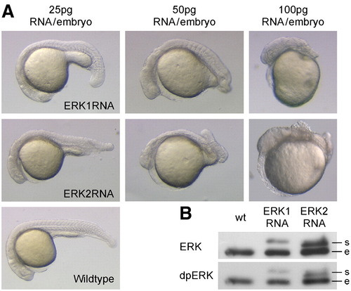
Over-expression of erk1 and erk2 led to concentration-dependent phenotypes. (A) Embryos were injected with 25, 50 or 100 pg erk1 RNA or erk2 RNA per embryo and developmental phenotypes were recorded at 24 hpf. (B) Western blot analysis was performed with global ERK and dpERK antibody on protein samples of 100 pg RNA injected embryos in shield stage. The proteins derived from synthetic mRNAs (s) are slightly larger than the endogenous proteins (e) due to the introduction of a small linker, and can therefore be distinguished by size. |
Expression Data
Expression Detail
Antibody Labeling
Phenotype Data
Phenotype Detail
Acknowledgments
This image is the copyrighted work of the attributed author or publisher, and
ZFIN has permission only to display this image to its users.
Additional permissions should be obtained from the applicable author or publisher of the image.
Reprinted from Developmental Biology, 319(2), Krens, S.F., He, S., Lamers, G.E., Meijer, A.H., Bakkers, J., Schmidt, T., Spaink, H.P., and Snaar-Jagalska, B.E., Distinct functions for ERK1 and ERK2 in cell migration processes during zebrafish gastrulation, 370-383, Copyright (2008) with permission from Elsevier. Full text @ Dev. Biol.