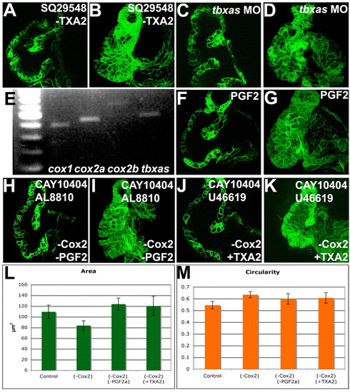
Activation of TXA2 signaling or inhibition of PGF2αrescued the Cox2 inhibition phenotype. Zebrafish embryos were treated with 10 μM SQ29548 (A,B), a TP receptor inhibitor, 25 μM CAY10404 and 5 μM AL8810 (H,I), an FP receptor inhibitor, 25 μM CAY10404 and 10 μM U46619 (J,K), a TP receptor agonist, or injected with tbxas morpholinos (C,D), imaged with confocal microscopy (A-D,F-K), projections made (B,D,G,I,K) and the area (L) and circularity (M) measured and the 95% confidence interval calculated. Myocardial bending (A,C,F) and cell shape changes (B,D,G) were observed in larvae in which TXA2 signaling was inhibited (A-D) or PGF2α signaling was increased (F,G). (E) RT-PCR was performed on RNA from isolated hearts for cox1, cox2a, cox2b and tbxas. cox1, cox2a, cox2b and tbxas are expressed in the heart. (H-M) Inhibition of PGF2α signaling (H,I) or activation of TXA2 signaling (J,K) rescued the Cox2 inhibition phenotypes of myocardial bending (H,J) and the decrease in myocardial cell area (I,K,L), but not the increase in myocardial circularity (M).
|

