
Fig. 4
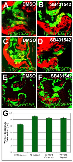
Inhibition of TGFβ signaling disrupted the shape of invaginating valve cells. (A-G) Zebrafish embryos were treated with 1% DMSO (A,C,E) or 3 μM SB431542 (B,D,F) from either 56-72 (A,B,E,F) or 56-96 (C,D) hpf, and stained with rhodamine-phalloidin (A-D) or for Alcam (E,F) and imaged by confocal microscopy (A-F) or SPIM (G). The thickness of the valve at the point of greatest compression or expansion was measured and compared along with the 95% confidence interval (G). (A-D) Inhibition of TGFβ signaling resulted in cuboidal invaginating cells (B,D) compared with control valves (A,C). (E,F) TGFβ signaling was required for the downregulation of Alcam in the invaginating cells. (G) This change in shape and adhesion resulted in the inability of the valves to compress and expand. |