
Fig. 7
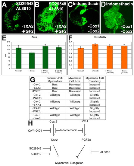
A balance of TXA2 and PGF2α signaling regulates myocardial cell shape. Zebrafish embryos were treated with 10 μM SQ29548 and 5 μM AL8810 (A,B), or 15 μM indomethacin (C,D), an inhibitor of Cox1 and Cox2, imaged with confocal (A-D), projections made (B,D) and the area (E) and circularity (F) measured and the 95% confidence interval calculated. Inhibition of both PGF2α and TXA2 signaling (A,B) or all Cox signaling (C,D) gave wild-type shape of the myocardium overlying the AVC (A,C) and wild-type myocardial area (B,D,E), although myocardial circularity was increased (F). (G) Table of the effects of inhibiting or activating different signaling pathways. (H) These findings suggest a model in which Cox2-derived TXA2 supports myocardial elongation, whereas PGF2α arising from Cox1 action or from the action of both Cox1 and Cox2 inhibits myocardial elongation. |