
Fig. 3
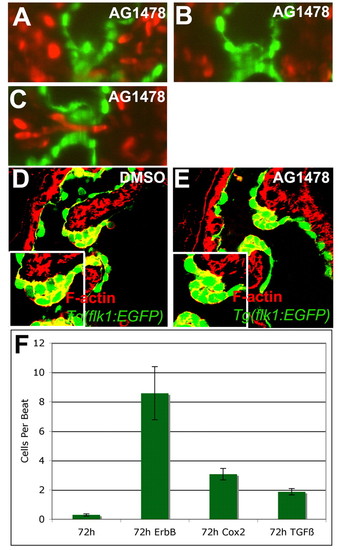
Inhibiting ErbB signaling disrupted the rolling of the valve. Zebrafish embryos were treated with 4 μM AG1478, an ErbB receptor inhibitor, or 1% DMSO from 56-72 hpf and imaged with SPIM (A-C) or stained with rhodamine-phalloidin and imaged with confocal microscopy (D,E). Retrograde flow of blood cells per beat was quantified for animals treated with 1% DMSO, 4 μM AG1478, 25 μM CAY10404 or 3 μM SB431542 along with the 95% confidence interval (F). (A-C) Inhibition of ErbB signaling led to altered valve function, with the valve coming together on the ventricular side of the AVC (A), rolling ventricularly to atrially (B), and coming apart during the relaxation phase allowing retrograde blood flow (C,F). Larvae treated with inhibitor showed malformed valves with incomplete invagination and increased space between the two sides of the valve leaflet (E). |