
Fig. 5
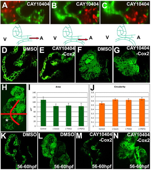
Inhibition of Cox2 and prostaglandin signaling altered myocardial cell shape. Zebrafish embryos were treated with 25 μM CAY10404 (A-C,E,G,M,N), a Cox2 inhibitor, or 1% DMSO (D,F,K,L) from 56-72 (A-C,D-G) or 56-60 (K-N) hpf, imaged using SPIM (A-C) or confocal microscopy (D-G,K-N) and projections made (F,G,L,N). The area (I) and circularity (J) of cells in the posterior outer curvature of the ventricle was measured and the 95% confidence interval calculated. (A-C) Inhibition of Cox2 caused the invaginating cells to be translated towards the ventricle, such that the AVC closed (A) and rolled (B) on an endocardial monolayer leading to retrograde blood flow (B). (D,E) The invaginating cells shifted towards the ventricle because the myocardium overlying the superior AVC bent towards the ventricle (E). (F,G) This bending correlates with changes in ventricular shape caused by decreases in cell area (G,I) and increases in cell circularity (G,J). (H) The ventricle was divided into four areas based on anterior or posterior, outer or inner curvature, for the cell shape and circularity analysis. Significant differences were found in the posterior outer curvature of the ventricle, indicated with an asterisk. Myocardial bending (M) and cell shape changes (N) arose within 4 hours of treatment with Cox2 inhibitor. |