
Fig. 5
- ID
- ZDB-FIG-080311-12
- Publication
- Vinothkumar et al., 2008 - Sequential and cooperative action of Fgfs and Shh in the zebrafish retina
- Other Figures
- All Figure Page
- Back to All Figure Page
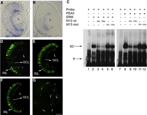
Pea3 and Erm regulate expression in the retina. (A, B) Erm (A) and Pea3 (B) are expressed at low levels throughout the retina with higher levels in the GCL at 34 hpf. (C) In vitro binding of Erm and Pea3 to the RetE probe (lanes 2, 8) and competition using M13 wild-type and M13 mutant oligos (lanes 3–6, 9–12) (SC, shifted complex; P, free probe). (D–G) Transgenic embryos (48 hpf) injected with either MO-pea3 (1.0 μg/μl) or MO-erm (1.0 μg/μl) had no effect on the expression pattern (E, F) while those injected with a mixture of both morpholinos (combined 1.0 μg/μl morpholino) showed a complete loss of GFP expression in the retina (G). Transgenic embryos injected with mismatch morpholinos show normal GFP expression in the GCL and INL (F). Anterior is to the top in all images. L, lens; GCL, ganglion cell layer; INL, inner nuclear layer. Scale bar, 50 μm. |
| Genes: | |
|---|---|
| Fish: | |
| Anatomical Terms: | |
| Stage: | Prim-15 |
Reprinted from Developmental Biology, 314(1), Vinothkumar, S., Rastegar, S., Takamiya, M., Ertzer, R., and Strähle, U., Sequential and cooperative action of Fgfs and Shh in the zebrafish retina, 200-214, Copyright (2008) with permission from Elsevier. Full text @ Dev. Biol.