
Fig. 2
- ID
- ZDB-FIG-080312-3
- Publication
- Vinothkumar et al., 2008 - Sequential and cooperative action of Fgfs and Shh in the zebrafish retina
- Other Figures
- All Figure Page
- Back to All Figure Page
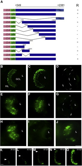
Deletion mapping of the retinal enhancer (RetE). A series of deletion constructs were generated (A) to identify the region required for shh expression in the retina. Transgenes carrying the region from + 549 to + 2381 and + 549 to + 2020 drive expression in the GCL and INL (B, C). The transgene carrying only the conserved region as ar-A from + 2021 to + 2381 failed to show any GFP expression in the retina (D). Transient expression of deletion constructs (E–J). The constructs carrying shh sequence from + 729 to + 2020 and + 549 to + 1035 mediate expression in the retina (E, F). The construct containing shh sequences from + 549 to + 829 did not give retina expression (G). Embryos injected with the -2.4shh:gfpRetE plasmid with the minimal region + 729 to + 1035 have GFP expression in the retina (H), while embryos injected with the -2.4shh:gfp + 549/2020 plasmid with an internal deletion from + 729 to + 1035 failed to drive expression in the retina (I). Embryos injected with the RetE (+ 549 to + 2020) in the context of a heterologous thymidine kinase (-37Tk) promoter -37tk:gfpRetE show retina expression (J). Anterior is to the top in all images and embryos were photographed at 72 hpf. (K–O) In vivo time lapse imaging of -2.4shh:gfpRetE expression in the retina. Single frames taken from a film recording of -2.4shh:gfpRetE expression in the retina, starting at 35 hpf and ending at 54 hpf. The spread of GFP expression is marked by arrowheads. R, retinal expression; L, lens; GCL, ganglion cell layer; INL, inner nuclear layer. Scale bar, 50 μm, panels B–J; 25 μm, panels K–O. +/- Indicates the presence or absence of GFP expression in the retina. |
Reprinted from Developmental Biology, 314(1), Vinothkumar, S., Rastegar, S., Takamiya, M., Ertzer, R., and Strähle, U., Sequential and cooperative action of Fgfs and Shh in the zebrafish retina, 200-214, Copyright (2008) with permission from Elsevier. Full text @ Dev. Biol.