
FIGURE
Fig. 3
- ID
- ZDB-FIG-080312-4
- Publication
- Vinothkumar et al., 2008 - Sequential and cooperative action of Fgfs and Shh in the zebrafish retina
- Other Figures
- All Figure Page
- Back to All Figure Page
Fig. 3
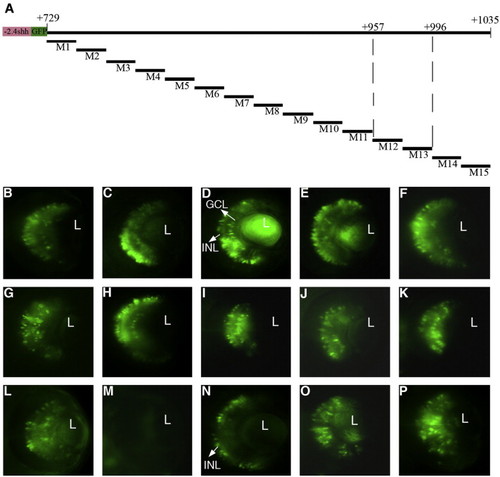
Fine mapping of the 300-bp RetE region. Non-overlapping clusters of point mutations of 20 bp were introduced into -2.4shh:gfpRetE from + 729 to + 1035 of the shh locus (A). The activity of mutant constructs M1–M15 was analyzed by transient expression (B–P). Clustered mutation M12 led to the complete loss of GFP expression (M) whereas mutation M13 abolished GFP expression exclusively in the GCL but not in the INL (N). All other mutant constructs show expression in both layers (B–L, O, P). L, lens; GCL, ganglion cell layer; INL, inner nuclear layer. Scale bar, 50 μm. |
Expression Data
Expression Detail
Antibody Labeling
Phenotype Data
Phenotype Detail
Acknowledgments
This image is the copyrighted work of the attributed author or publisher, and
ZFIN has permission only to display this image to its users.
Additional permissions should be obtained from the applicable author or publisher of the image.
Reprinted from Developmental Biology, 314(1), Vinothkumar, S., Rastegar, S., Takamiya, M., Ertzer, R., and Strähle, U., Sequential and cooperative action of Fgfs and Shh in the zebrafish retina, 200-214, Copyright (2008) with permission from Elsevier. Full text @ Dev. Biol.