
Fig. 1
- ID
- ZDB-FIG-080312-2
- Publication
- Vinothkumar et al., 2008 - Sequential and cooperative action of Fgfs and Shh in the zebrafish retina
- Other Figures
- All Figure Page
- Back to All Figure Page
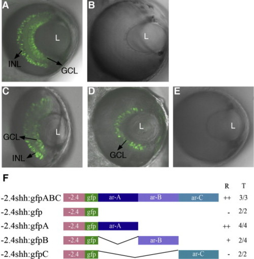
Expression patterns of shh transgenes in the retina. Transgenic lines -2.4shh:gfpABC and -2.4shh:gfpA drive expression in the ganglion cell layer (GCL) and inner nuclear layer (INL) of the zebrafish retina (A, C). In the -2.4shh:gfpB transgenic line, GFP expression is seen only in the GCL (D). The -2.4shh:gfp and -2.4shh:gfpC (B, E) transgenes do not drive GFP expression in the retina. ‘L’ denotes lens. Anterior is to the top in all images. Confocal images taken at 72 hpf. (F) Outline of the enhancer constructs. ‘R’ indicates the expression in the retina and ‘T’ the number of stable transgenic lines showing retina expression out of the total lines analyzed. Scale bar, 50 μm. ‘++’: Expression in both GCL and INL; ‘+’: expression in GCL; ‘-’ no GFP expression in retina. |
| Gene: | |
|---|---|
| Fish: | |
| Anatomical Terms: | |
| Stage: | Protruding-mouth |
Reprinted from Developmental Biology, 314(1), Vinothkumar, S., Rastegar, S., Takamiya, M., Ertzer, R., and Strähle, U., Sequential and cooperative action of Fgfs and Shh in the zebrafish retina, 200-214, Copyright (2008) with permission from Elsevier. Full text @ Dev. Biol.