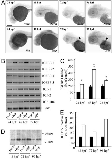
Hypoxia induces IGFBP-1 expression. (A) Hypoxia increased IGFBP-1 mRNA expression in many tissues in early embryonic stages (24 and 48 hpf) and in the liver (indicated by arrow heads) in advanced stages (72 and 96 hpf). Zebrafish embryos at various stages were transferred to normal or hypoxic water. After 24 h, the embryos were fixed and subjected to in situ hybridization analysis. (Scale bar, 200 μm.) (B) RT-PCR analysis of IGFBP-1, IGFBP-2, IGFBP-3, IGFBP-5, IGF-1, IGF-2, IGF-1 receptor-a (IGF-1Ra), and ornithinedecarboxylase (odf) in embryos of the indicated stages. Norm, normoxia; Hyp, hypoxia. (C) Relative IGFBP-1 mRNA levels normalized by the ornithinedecarboxylase mRNA level. Filled bars represent the normoxia group, and open bars represent the hypoxia group. Values are expressed as means ± SE. (n = 6). *, P < 0.05 compared with the corresponding normoxia group. (D) Western ligand blotting analysis. Wild-type embryos at various indicated stages were subjected to 24 h of hypoxia or normoxia treatment. The levels of IGFBP-1 protein were determined by ligand blotting with digoxigenin-labeled IGF-1. (E) Densitometric analysis results of D. Filled bars represent the normoxia group, and open bars represent the hypoxia group.
|
