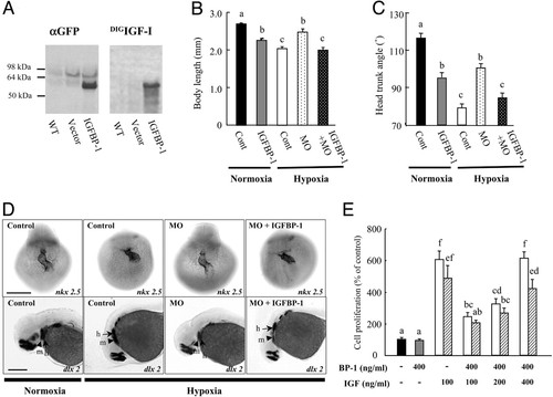
IGFBP-1 inhibits embryonic growth and development by inhibiting IGF actions. (A) Expression of a functional IGFBP-1 fusion protein. Embryos were injected with the IGFBP-1–EGFP construct (IGFBP-1) or empty vector (vector). At 36 hpf, the embryos were subjected to immunoblot (Left) and ligand blot analysis (Right). WT, wild-type embryos; vector, empty-vector-injected embryos; IGFBP-1, embryos injected with the IGFBP-1–EGFP construct. (B and C) Effect of IGFBP-1 overexpression under normoxia and hypoxia. Embryos injected with control MO (Cont), IGFBP-1 MO1 (MO), or IGFBP-1 MO plus IGFBP-1–EGFP overexpression vector at 12 hpf were transferred to normal (normoxia) or hypoxic water (hypoxia). The body length (B) and HTA (C) were measured 24 h later. Values are expressed as means ± SE (n = 20–34). Groups with common letters are not significantly different from each other (P < 0.05). (D) MO knockdown abrogates and IGFBP-1 overexpression restores hypoxia-induced delay in head skeleton and heart morphogenesis. Embryos injected with control MO, IGFBP-1 MO, or IGFBP-1 MO plus IGFBP-1–EGFP overexpression vector were transferred to normal or hypoxic water for 24 h and subjected to whole-mount in situ hybridization with nkx 2.5 (Upper) and dlx 2 (Lower). h, hyroid arch (arrows); m, mandibular arch (arrow heads). (Scale bar, 200 μm.) (E) IGFBP-1 inhibits IGF-stimulated zebrafish embryonic cell proliferation. Serum-starved, confluent cells were exposed to IGFBP-1 (400 ng/ml) with or without various concentrations of IGF-1 (open bars) or IGF-2 (shadow bars) in the presence of BrdUrd (20 μM) for 24 h. BrdUrd-labeled cells were detected by immunostaining. Values are expressed as means ± SE of two independent experiments, each of which was performed in duplicates. Groups with the same letters (a, b, c, d, e, or f) are not significantly different from each other (P < 0.05).
|
