
- Title
-
Zebrafish wnt9a is expressed in pharyngeal ectoderm and is required for palate and lower jaw development
- Authors
- Curtin, E., Hickey, G., Kamel, G., Davidson, A.J., and Liao, E.C.
- Source
- Full text @ Mech. Dev.
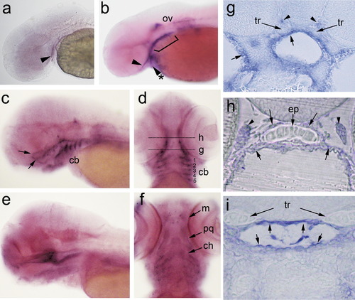
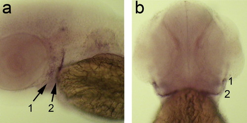
Expression of wnt9a in pharyngeal arch and oropharyngeal epithelium. Whole-mount RNA in situ hybridization of wnt9a at 36 hpf (a), 48 hpf (b), 3 dpf (c, d, g and h), and 4 dpf (e, f and i). Embryos are oriented with head toward the left in lateral views (a–c and e) and head toward the top in ventral views (d and f) and sections (g–i). Sections are shown in 10× (g), and 40× (h and i) magnification. The levels of the sections shown in panels (g) and (h) are indicated in panel (d), and the level in panel (i) is indicated in (e). The wnt9a transcripts can be detected in the developing pharyngeal arches (arrow, a) as paired cell clusters. At 48 hpf, wnt9a expression is detected in the first pharyngeal arch, as it splits to dorsal (b, arrow) and ventral (b, arrow with asterisk) regions. There is also wnt9a expression along the posterior pharyngeal arch cells (c, bracket), and the otic vesicle (ov). By 3 dpf, facial skeletal structures have formed, where wnt9a expression can be detected surrounding the ethmoid plate (ep), trabeculae (tr), and lower jaw structures, including the five paired ceratobrachials (cb, 1–5). To better visualize this weak whole-mount staining, plastic sections were performed (g and h). Cross-section analysis reveals wnt9a expression in the oral epithelium along the roof and the floor of the oropharynx (g, arrow), and along the surface epithelium ventral to the lower jaw (g, arrow with asterisk). The ethmoid plate is surrounded by wnt9a expressing cells, bordered by the roof of the oropharynx below, and a single cell-layer of wnt9a expressing cells above (h, arrows). There are also clusters of wnt9a expressing cells laterally that may represent ocular muscle or optic nerve structure (h, arrowheads). Expression of wnt9a persists into 4 dpf along the oropharynx and continues into the foregut (e), detected in the epithelium that lines the oropharynx circumferentially (i). There is also weak wnt9a expression outlining the skeletal structures, such as Meckel’s cartilage (m), palatoquadrate (pq) and the ceratohyal (ch). Abbreviations: cb, ceratobrachial; ch, ceratohyal; ep, ethmoid plate; m, Meckel’s cartilage; ov, otic vesicle; pq, palatoquadrate; tr, trabeculae. |
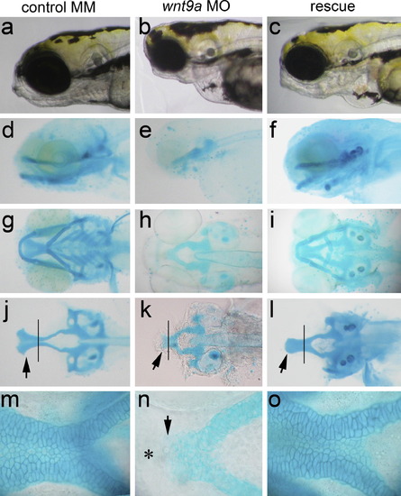
Knockdown of wnt9a profoundly disrupts craniofacial development. Live embryo morphology (a–c) and Alcian blue cartilage stain (d–o). Lateral view (a–f), ventral view (g–i), ventral view with lower jaw dissected away (j–l), taken at 10× magnification. View of the ethmoid plate of embryos in (g–i) at 40× magnification (m–o). Anterior is to the left in all views. Embryos injected with wnt9a mismatch control (MM) (a, d, g, j and m), translation-blocking morpholino (MO) (b, e, h, k and n), and wnt9a MO with wnt9a full-length capped mRNA rescue at 4 dpf (c, f, i, l and o). Normal craniofacial morphology (a) and jaw structures are seen in embryos injected with mismatch-control morpholino, with normal lower jaw (g) and ethmoid plate (j, arrow) and lower jaw (arrow in d, j). Knockdown of wnt9a produced loss of jaw structure apparent by facial morphology (b). In the wnt9a morphant, Alcian blue staining detects formation of the medial parachordals but the lateral parachordal structures are absent (h and k). In the wnt9a morphant, the proximal trabeculae form and converge in midline but fail to elongate, and the ethmoid plate does not form (arrow, k, n), and the lower jaw is absent. When wnt9a MO is co-injected with wnt9a mRNA, the morphant phenotype is rescued, albeit not entirely wild-type (c, f, i, l and o). In the rescued animals, the ethmoid plate forms but the trabelculae fail to flair out in the distal leading edge (arrow, l). The lower jaw and the parachordals form in the rescued embryos, but the vertex of the ceratohyal points posteriorly, and the ceratobrachials are not formed. Higher magnification comparison of the trabeculae between the wnt9a morphant and the controls show that the morphant chondrocytes fail to adopt the elongated morphology and remain rounded, with abrupt cutoff anteriorly, as the trabeculae end in a stump (arrow, n). There are no cells found anterior to the cutoff (asterisk). PHENOTYPE:
|

Analysis of cranial neural crest cells after wnt9a knockdown in sox10:eGFP transgenics. Embryos injected with MM control (a, c and e) and translation-blocking MO (b, d and f), where the cranial neural crest cells (CNCC) are followed over embryogenesis. Embryos are shown in ventral views at 48 hpf (a and b), 72 hpf (c and d) and 96 hpf (e and f). Initial CNCC delamination from the neural plate occurs normally in the MO injected embryos (not shown) and the first distinct phenotype is observed with when the dorsal group of CNCC that differentiate into the trabeculae become obliquely oriented and the distal leading edge migrate toward the midline (arrows, a). In the MO injected embryos, the dorsal CNCC fail to move medially, resulting in a wide separation between the CNCC groups. At 72 hpf, the ethmoid plate and the trabeculae are formed (c). In the morphant, the trabeculae is formed but does not join in the midline, and the ethmoid plate is absent (d). By 96 hpf, the distal edges of the morphant trabeculae come into the midline and is joined by 1–2 cell layers of tissue, with absent ethmoid plate (f). Abbreviations: ep (ethmoid plate), tr (trabeculae). |
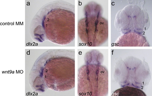
Knockdown of wnt9a does not alter early cranial neural crest migration. Embryos injected with wnt9a mismatch control (MM) (a–c), and translation-blocking morpholino (MO) (e–h). Timepoints are 24 hpf (a, b, d and e), and 42 hpf (c and f). Embryos are mounted in lateral view with head toward left detected with dlx2a riboprobe (a and d), detected with sox10 riboprobe in dorsal view with head toward top (b and e), and gsc riboprobe in ventral view (c and f). These neural crest markers all demonstrate normal early migration to appropriate pharyngeal arch positions, in mismatch (MM) and translation-blocking (MO) morpholino injected embryos. No developmental delay was observed. |
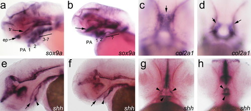
Knockdown of wnt9a results in late craniofacial phenotypes. Embryos injected with wnt9a mismatch control (MM) (a, c, e and g), and translation-blocking morpholino (MO) (b, d, f and h). Embryos are mounted in lateral view with head toward left (a, b, e–h) and in ventral view with head toward top (c and d). Transcripts of sox9a (a and b) at 3 dpf, col2a (c and d) at 2 dpf, and shh (e–h) at 2 dpf are detected by whole mount RNA in situ hybridization. The truncated trabeculae and absent ethmoid plate are confirmed by comparison of sox9a expression between MO (b) and MM control (a). In the wnt9a morphant, sox9a expressing cells do not elongate to form the trabeculae, and there is no ethmoid plate (b, arrow). The expression of sox9a in the posterior pharyngeal arches is also absent (b, bar). The expression of col2a is present but aberrantly located in the wnt9a morphant, distinguishable from MM control as early as 48 hpf. In the MM control, col2a expression prefigures the trabeculae and the ethmoid plate (c), whereas in the morphant, the cranial neural crest cells are lateralized in linear groups (d). Oral epithelium express shh in a wildtype pattern in the MM control and the morphant, outlining the stomodeum (g, arrowheads). The stomodeum forms in the wnt9a morphant, although the shape is altered (f, arrowheads), and is continuous to the foregut, with shh expression outlining the roof (e, f, arrows) and floor (e, f, arrowheads) of the oropharynx. EXPRESSION / LABELING:
PHENOTYPE:
|

RT-PCR for wnt9a transcript in somite-stage time points. Transcripts of wnt9a are detectable via RT-PCR from as early as 5-somite stage. However, by whole mount RNA in situ, the first tissue-restricted expression of wnt9a is not apparent until 36 hpf. EXPRESSION / LABELING:
|

Expression of wnt9a in the pronephric duct. The developing pronephric duct expresses wnt9a, from d4 onward. Expression is detected from the start of the pronephric duct in the embryonic renal anlage, running the length of the collecting tubule (arrows) and extends to the cloaca (arrowhead). EXPRESSION / LABELING:
|
Expression of wnt9b in pharyngeal arches. Whole mount RNA in situ analysis of wnt9b at 48hpf, lateral view (a) and ventral view (b). Discrete wnt9b expression is detected in cells populating pharyngeal arches 1 and 2 (arrows), as previously reported. <EXPRESSION / LABELING:
|
Reprinted from Mechanisms of Development, 128(1-2), Curtin, E., Hickey, G., Kamel, G., Davidson, A.J., and Liao, E.C., Zebrafish wnt9a is expressed in pharyngeal ectoderm and is required for palate and lower jaw development, 104-115, Copyright (2011) with permission from Elsevier. Full text @ Mech. Dev.