
- Title
-
Mutations affecting neurogenesis and brain morphology in the zebrafish, Danio rerio
- Authors
- Jiang, Y.J., Brand, M., Heisenberg, C.P., Beuchle, D., Furutani-Seiki, M., Kelsh, R.N., Warga, R.M., Granato, M., Haffter, P., Hammerschmidt, M., Kane, D.A., Mullins, M.C., Odenthal, J., van Eeden, F.J., and N�sslein-Volhard, C.
- Source
- Full text @ Development
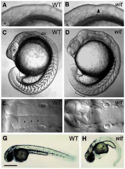
Phenotype of live witta52b embryos. If not specified, all the following figures are rostral to the left, dorsal to the top. Lateral views of wild-type (A) and mutant (B) embryos at 9-somite stage. The indentation in the hindbrain (arrowhead) is indicated. Lateral views of wild-type (C) and mutant (D) embryos at 17-somite stage. The indistinct somite boundaries (bracket) are indicated. Dorsal views of wild-type (E) and mutant (F) embryos at 26 hours postfertilization (hpf). Normal rhombomeric boundaries (arrowheads) are not seen in mutant embryos. Lateral views of wildtype (G) and mutant (H) embryos at 36 hpf. Note that there are no visible pigmented melanophores in the posterior trunk of mutant embryo in H. e, eye; ov, otic vesicle. Bar, 200 �m (A,B,E,F); 335 �m (C,D); 640 �m (G,H). PHENOTYPE:
|

Neuronal hyperplasia in witta52b. All are dorsal views and rostral to the left, except for G and H, which are lateral views and rostral to the top. Hindbrain reticulospinal neurons in wild-type (A) and mutant (B) embryos labeled with mAb 3A10 at 36 hpf. Anterior parts of wild-type (C) and mutant (D) embryos and of trunk parts of wild-type (E) and mutant (F) embryos labeled with Islet-1 antibody at the 8-somite stage. Wildtype (G) and mutant (H) embryos labeled with Islet-1 antibody at 20-somite stage. The presumptive anterior lateral line ganglion (arrowhead) and presumptive posterior lateral line ganglion (arrow) are indicated. pax-b expression in wild-type (I) and mutant (J) embryos at 24 hpf. The CoSA neurons (arrowheads) and pronephric duct (arrows) are indicated (Mikkola et al., 1992). In mutant embryos, not only is the pax-b expression in spinal interneurons and pronephric duct increased, and there is also an increased expression in presumptive r2, r3 and r4 (asterisks). dr, dorsorostral cluster; e, epiphysis; M, Mauthner cell in r4; mhb, midbrain-hindbrain boundary; mlf, medial longitudinal fascicle; mn, primary motoneurons; op, otic placode; os, optic stalk; ov, otic vesicle; p, polster; rb, Rohon-Beard cells; RoL2, RoL2 neurons in r2; tg, trigeminal ganglia; vr, ventrorostral cluster. Bar, 100 �m (A,B); 160 �m (CF); 255 �m (G,H); 400 �m (I,J). EXPRESSION / LABELING:
PHENOTYPE:
|

The expression pattern of two zebrafish achaete-scute homolog genes in white tail. Zash-1a expression in wild-type (A) and mutant (B) embryos at the 17-somite stage. The presumptive mhb (arrowhead) and presumptive r3 (asterisk) are indicated. Zash-1b expression in wild-type (C) and mutant (D) embryos at 15-somite stage. The expression in the spinal cord (triangles) is indicated. Wildtype (E) and mutant (F) embryos at 26 hpf labeled with Zash-1a RNA probe. Wildtype (G) and mutant (H) embryos at 26 hpf labeled with Zash- 1b RNA probe. ov, otic vesicle; r(n), rhombomere (n). Bar, 320 �m (A-D); 100 �m (E-H). EXPRESSION / LABELING:
|
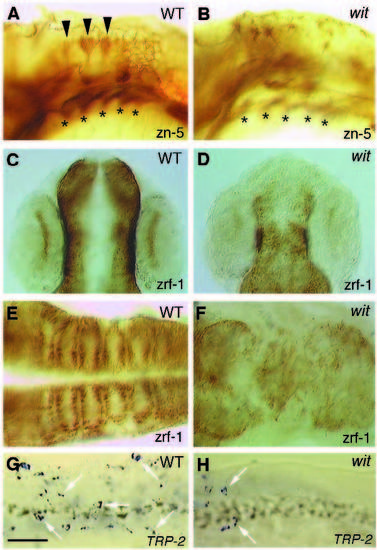
Some cell types are decreased in witta52b. C and D are rostral to the top. A, B are lateral views; C-H are dorsal views. Wildtype (A) and mutant (B) embryos labeled with zn-5 mAb at 36 hpf. The dorsal hindbrain neurons (arrowheads; Trevarrow et al., 1990) are decreased in mutant embryos. The pharyngeal endoderm (asterisks; Schilling and Kimmel, 1994) is not affected. The pharyngeal endoderm in B is out of focus. Wildtype (C,E) and mutant (D,F) embryos labeled with zrf-1 mAb. C,D are at 24 hpf; E,F are at 38 hpf. Radial glial fibers in the retina (Westerfield, 1994) and hindbrain (Trevarrow et al., 1990) are decreased. TRP-2 expression pattern of wild-type (G) and mutant (H) embryos in the anterior trunk at 24 hpf. The labeled melanophores (arrows) are decreased in H. Bar, 125 �m (A-D); 100 �m (E-H). |

Somitogenesis is delayed, but somitic myogenesis is normal in witta52b. A,B,E,F dorsal views. C,D lateral views. Myf-5 antibody labeling in wild-type (A) and mutant (B) embryos at the 11-somite stage. MF20 labeling in wild-type (C) and mutant (D) embryos at the 17-somite stage, rostral to the top. The somite boundaries (arrowhead) are indicated. Notch expression in wild-type (E) and mutant (F) embryos at the 12-somite stage. psm, presomitic mesoderm. Bar, 400 �m (A,B); 240 �m (C,D); 475 �m (E,F). EXPRESSION / LABELING:
PHENOTYPE:
|

Phenotype of live pac embryo. C-E treated with 0.2 mM PTU. Lateral views of wild-type (A) and pactj250a (B) embryos at around the 9-somite stage. The small bulge (arrowhead) in the hindbrain is indicated. Lateral views of wild type (C) at 28 hpf, pactm101b (D) at 28 hpf and pactg1a (E) at 36 hpf. The filopodia-like structure (arrow) is indicated. Bar, 320 �m (A,B); 200 �m (C-F). PHENOTYPE:
|

Disorganization of the hindbrain in pac. Dorsal views of wild type (A) and pactj250a (B) labeled with zrf-1 mAb. Dorsal views of wild type (C) and pactj250a (D) labeled with mAb against acetylated tubulin. Bar, 125 �m. |

Variable disorganization of hindbrain reticulospinal interneurons in pactj250a at 36 hpf shown by 3A10 mAb staining; dorsal views with rostral to the top. In wild type (A), the most prominently labeled Mauthner cells are located in r4, which project axons contralaterally to the mlf. RoL2 exists in r2, which projects axons contralaterally to the mlf, then to the llf. In A, B and C, MiM1(or MiV1) (arrowheads; Mendelson, 1986a, b) is indicated. According to their position and axonal projection, the two neurons in r6 could be MiD3i (asterisk), which has an ipsilateral axon and MiD3c (triangle), which has a contralateral axon, as shown in C. B, C and D are representatively labeled embryos. In most of the cases, as shown in B, one of the Mauthner cells in rhombomere 4 is displaced to a more posterior rhombomere (r5, r6 or in between) and locates in the same side of the normal one (n=5/10). In B, a presumptively displaced MiD3c (arrow) is indicated. In a few cases, as shown in C, the displaced Mauthner cell locates in the opposite side (n=1/10). Sometimes, as shown in D, both of the Mauthner cells are displaced to a more posterior rhombomere, and their axons project to the ipsilateral instead of the contralateral side (n=3/10). llf, lateral longitudinal fascicle; M, Mauthner cell; mlf, medial longitudinal fascicle; ov, otic vesicle; pllg, posterior lateral line ganglion; RoL2, RoL2 neurons in r2; tpll, tract of posterior lateral ganglion. Bar, 125 �m. |
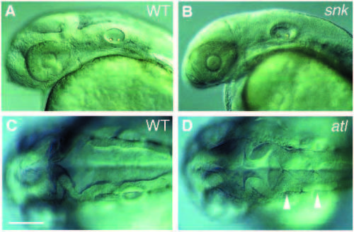
Phenotypes of live snktw3b and atltc234b. Lateral views of wild type (A) and snktw3b (B) at 36 hpf, treated with 0.2 mM PTU. Dorsal views of wild-type (C) and atltc234b (D) embryos at 24 hpf. The extra neural folds (arrowheads) are indicated. Bar, 250 �m (A,B); 200 �m (C,D). |
