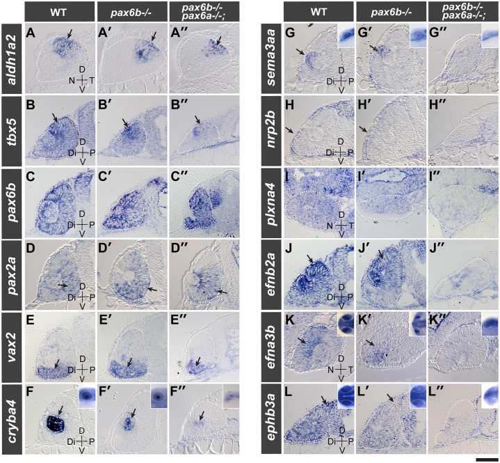
Image
Figure Caption

Fig 8 Pax6a/b compound mutants lack expression of guidance molecules in the eye.

Gene expression was analysed by in situ hybridisation in wild type (WT, A-L), maternal zygotic pax6b-/- (A’-L’) and pax6a-/-;pax6b-/- double homozygous mutant embryos (A”-L”) at 28 hpf. The gene symbol of the transcript analysed is given at the left end of each row. The stippled white line demarcates the optic cup and brain. Sections are made either in transverse orientation (tbx5 [B-B”], pax6b [C-C’], pax2a [D-D”], vax2 [E-E”], cryba4 [F-F”], sema3aa [G-G”], nrp2b [H-H”], efnb2a [J-J”] and ephb3a [L-L”]) or in sagittal orientation (aldh1a2 [A-A”], plxna4 [I-I”] and efna3b [K-K”]) to show the gene expression along the distal-proximal and nasal-temporal axis, respectively. For cryba4 (F-F”), sema3aa (G-G”), efna3b (K-K”) and ephb3a (L-L”), the head region of the whole mount embryos is shown in the inset. Note the expression indicated by the arrow: dorso-temporal for aldh1a2 (A-A”), dorsal for tbx5 (B-B”), ventro-proximal for pax2a (D-D”), ventral part of the optic cup for vax2 (E-E”), lens primordium for cryba4 (F-F”) and sema3aa (G-G”), distal periocular mesenchymal cells for nrp2b (H-H”), dorsal for efnb2a (J-J’), dorso-nasal optic cup for efna3b (K-K”) and periocular mesenchymal cells for ephb3a (L-L”). Scale bar: 50 μm; 200 μm for insets.

Figure Data
Acknowledgments
This image is the copyrighted work of the attributed author or publisher, and ZFIN has permission only to display this image to its users. Additional permissions should be obtained from the applicable author or publisher of the image. Full text @ PLoS Genet.