
Figure 12.
- ID
- ZDB-FIG-221211-320
- Publication
- Nelson et al., 2022 - The developmental progression of eight opsin spectral signals recorded from the zebrafish retinal cone layer is altered by the timing and cell type expression of thyroxin receptor β2 (trβ2) gain-of-function transgenes
- Other Figures
- All Figure Page
- Back to All Figure Page
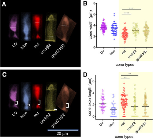
Thyroxin-receptor-β2 gain-of-function transgenes alter cone morphology. A, Width of cone types identified by transgene markers, is measured at the greatest extent of the inner segment. B, The trβ2 gain-of function cones are significantly wider than red-cones. C, The cone axon length is measured from the base of the inner segment to the apex of the cone pedicle. D, The trβ2 gain-of-function axon lengths are significantly shorter than red-cone axon lengths. B, D, Asterisks indicate significant differences (GraphPad convention, ANOVA and Tukey post hoc p-values given in text). A, C, Images are of 6-dpf in vivo larval fluorescent cones from confocal stacks. Control UV and red cones were imaged in sws1:GFP;trβ2:tdTomato larvae; blue cones, in sws2:GFP larvae; fluorescent trβ2 gain-of-function cones, in crx:mYFP-2A-trβ2 and gnat2:mYFP-2A-trβ2;mpv17−/− larvae. Larvae were anesthetized with MS222 and embedded in agarose after raising to 6 dpf in 300 μm PTU to block melanin formation in the pigment epithelium. |