
Figure 3.
- ID
- ZDB-FIG-221211-311
- Publication
- Nelson et al., 2022 - The developmental progression of eight opsin spectral signals recorded from the zebrafish retinal cone layer is altered by the timing and cell type expression of thyroxin receptor β2 (trβ2) gain-of-function transgenes
- Other Figures
- All Figure Page
- Back to All Figure Page
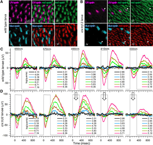
Opsin distributions and spectral responses in embryonic wild-type and crx:mYFP-2A-trβ2 larval eyes. Ai, UV opsin (SWS1) immunoreactive cones in a WT retina. Aiii, Avi, Red opsin (LWS1, LWS2) immunoreactive cones in WT retinas. Aiv, Blue-opsin (SWS2) immunoreactive cones in a WT retina. Aii, UV and red opsins are expressed in separate cones in a WT retina. Av, Red and blue opsins are expressed in separate cones in a WT retina. Bi, UV-opsin immunoreactive cones in a crx:trβ2 retina. Biii, Bvi, Red-opsin immunoreactive cones in crx:trβ2 retinas. Biv, Blue-opsin immunoreactive cones in a crx:trβ2 retina. There are fewer UV and blue cones in crx:trβ2 retinas than in WT retinas. Bii, One crx:trβ2 cone is immunoreactive for both UV and red opsins (arrowhead). Bv, Arrowhead points to a crx:trβ2 cone immunoreactive for both red and blue opsins. C, Cone signals from a WT larval eye respond to all stimulus wavelengths with largest amplitudes at 490 nm. D, A larval crx:trβ2 retina responds with maximal amplitudes at wavelengths 490, 570, and 650 nm but is less responsive than WT for 330, 410, and 490 nm (arrows), wavelengths that stimulate blue and UV cones. A, B, 5-dpf larvae. C, D, 6-dpf larvae. Perfusion medium contains 20 mm aspartate to isolate photoreceptor signals in the ERG. Five of nine stimulus-protocol wavelengths are illustrated. The stimulus irradiances [units of log(quanta·μm−2·s−1)] appear in legends to the right of stacked irradiance-response traces. |