
Fig. 5-S1
- ID
- ZDB-FIG-180417-26
- Publication
- Rogers et al., 2017 - Nodal patterning without Lefty inhibitory feedback is functional but fragile
- Other Figures
- All Figure Page
- Back to All Figure Page
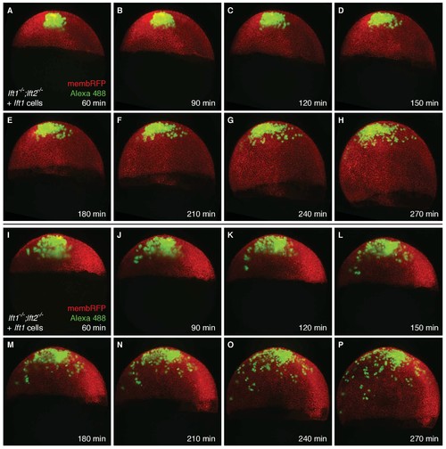
Transplanted cells are mostly retained in the animal pole during germ layer patterning. lft1-/-;lft2-/- mutant donors were injected with 0.4 pg Alexa 488-dextran (3 kD, ThermoFisher), 10 pg lft1 mRNA, and 30 pg membrane RFP mRNA at the one-cell stage; lft1-/-;lft2-/- mutant hosts were injected with 30 pg membrane RFP mRNA. At sphere stage, around 50 cells from donors were transplanted into the animal pole of host embryos as in Figure 5A,E and F. Embryos were imaged using a Zeiss Lightsheet Z.1 microscope starting 60 min post-transplantation for 3.5 hr. Maximum intensity projections (~525 µm total) are shown for two embryos (A–H and I–P); time post-transplantation is indicated in the lower right corner of each image. Transplanted cells tended to stay mainly in the animal pole during the imaging session, when the majority of Nodal-mediated germ layer patterning occurs (late blastula - early gastrula stages [Hagos and Dougan, 2007]). This supports the idea that ectopic Lft expression far from its endogenous source at the margin is sufficient to generate viable embryos (see Figure 3—figure supplement 1A,B for endogenous lft expression domains). Note that due to constant production of membrane RFP, the minimum and maximum display values for the red channel were manually adjusted in each frame. |