
Fig. 4
- ID
- ZDB-FIG-180208-14
- Publication
- Donat et al., 2018 - Heg1 and Ccm1/2 proteins control endocardial mechanosensitivity during zebrafish valvulogenesis
- Other Figures
- All Figure Page
- Back to All Figure Page
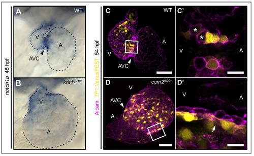
Notch activity within endocardial cushion cells is affected in ccm mutants. (A, B) Whole-mount in situ hybridization of notch1b cardiac expression at 48 hpf. (A) notch1b expression is restricted to endocardial cells of the atrioventricular canal (AVC) in wild-type (WT), while notch1b is strongly expressed throughout all ventricular cells in krit1ty219c mutants (B). (C, D) Projections of single confocal z-section images of endocardial cells marked by Tg(TP1:VenusPEST)s940 expression (yellow) and Alcam staining (magenta) at 54 hpf. (C) In WT, Notch activity is highest in endocardial cells of the AVC and outflow regions. (C') Single confocal plane section of the AVC (white box in C) reveals that some endocardial cells close to the ventricle lack Notch activity (asterisks). (D) In ccm2m201 mutants, the domain of high Notch activity is expanded to most ventricular endocardial cells. (D') Single confocal plane section of the AVC region (box in D, arrow) shows high Notch expression in all endocardial cells of the AVC region. A: atrium, V: ventricle, Scale bars are 50 μm (C, D), and 10 µm (C', D'). |
| Genes: | |
|---|---|
| Antibody: | |
| Fish: | |
| Anatomical Terms: | |
| Stage: | Long-pec |
| Fish: | |
|---|---|
| Observed In: | |
| Stage: | Long-pec |