
Fig. 3 S4
- ID
- ZDB-FIG-180208-12
- Publication
- Donat et al., 2018 - Heg1 and Ccm1/2 proteins control endocardial mechanosensitivity during zebrafish valvulogenesis
- Other Figures
- All Figure Page
- Back to All Figure Page
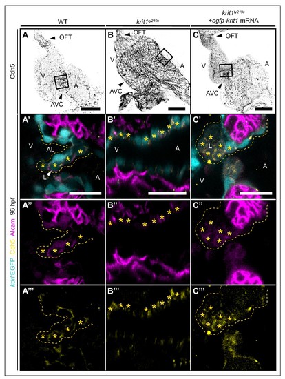
Cadherin-5 is misexpressed in krit1ty219c mutant embryonic hearts. (A–C) Projections of confocal z-stack images of 96 hpf embryonic hearts marked by Cadherin 5 (Cdh5) staining. (A'–C''') Details are shown in single confocal plane sections of the atrioventricular canal (AVC) region (boxes) of superior cardiac valve leaflets of 96 hpf hearts marked by Tg(kdrl:EGFP)s843 expression (cyan), Cdh5 staining (yellow), and Alcam staining (magenta). (A'–A''') At 96 hpf, Alcam and Cdh5 expression is mainly restricted to luminal endocardial cells of the developing wild-type (WT) cardiac valve leaflet (asterisks). (B'–B''') krit1ty219c mutants do not form valve leaflets and endocardial cells express Alcam and Cdh5 (asterisks). (C'–C''') Injection of egfp-krit1 mRNA into krit1ty219c mutants results in a dysmorphic cardiac valve leaflet at 96 hpf with an agglomeration of luminal leaflet cells marked by Alcam and Cdh5 expression (asterisks) (n = 17/23 krit1ty219c mutants). A: atrium, V: ventricle, L: luminal, AL: abluminal. Scale bars are 50 μm (A–C) and 20 µm (A'–C'''). |