|
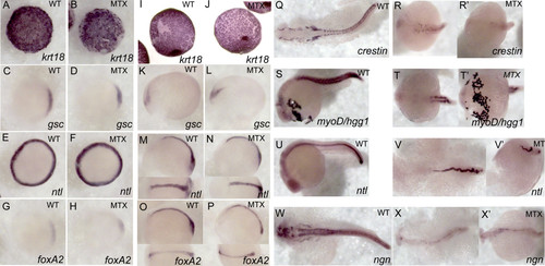
Methotrexate–treated embryos maintain expression of appropriate developmental markers. Expression patterns were visualized via whole-mount in situ hybridization in embryos treated with 400 μM MTX and fixed at shield stage (A-H), tailbud (I-P), and 24 hpf (Q-W′). While MTX-treated embryos at 24hpf are clearly delayed with respect to normal development, they do express appropriate developmental markers: krt18 –enveloping layer, gsc – involuting cells and neural plate, ntl – blastomere margin, notochord and dorsal organizer, foxA2 – involuting cells and neural plate, crestin – neural crest, myoD – paraxial mesoderm, hgg- hatching gland (anterior most structure), ngn – CNS. (A-J) Animal view, dorsal to the right. (K-P) Lateral view with dorsal view inset. (Q, R, R′, T, V, W, X, X′) Dorsal view. (S, U, V′) Lateral view. (T′) ventral view.
|