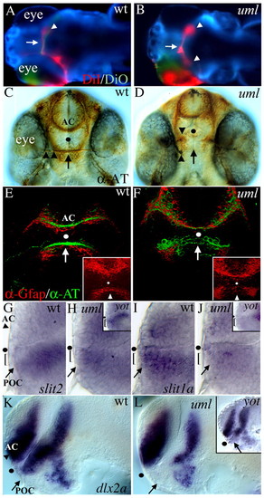
Axon guidance and forebrain patterning defects in umleitung (uml) mutants. (A,B) DiI (red) and DiO (green) labeling of retinal ganglion cell (RGC) axons from the left eye at 5 days postfertilization (dpf). Dorsal views, anterior to the left. In wild-type zebrafish (A), all RGC axons cross the midline (arrow) to innervate the contralateral tectal lobe (arrowhead). In uml mutants (B) some RGC axons fail to cross the midline (arrow) and instead project to the ipsilateral tectal lobe, leading to bilateral projections (arrowheads). (C,D) Anti-acetylated tubulin (AT) labeling of forebrain axons at 48 hpf. Ventral views, anterior uppermost. In wild-type embryos (C), axons cross the midline to form the anterior commissure (AC) in the telencephalon and the post optic commissure (POC, arrow) in the diencephalon. RGC axons (arrowheads) grow along the POC (arrow) to the midline to form the optic nerve and optic chiasm. In uml mutants (D), RGC axons fail to reach the midline at this age (arrowheads) and the POC fails to form (arrow), whereas the AC forms normally in the telencephalon. (E,F) Anti-Gfap (red) and anti-AT (green) labeling of glia and axons in the forebrain at 36 hpf. Ventral views, anterior at the top. Insets show Gfap labeling alone. In wild-type embryos (E), glial cells span the midline adjacent to the AC and POC (arrow). In uml mutants (F), the posterior glial bridge is disrupted, leaving a gap at the midline (arrow). (G-L) In situ hybridization (ISH) showing gene expression in the anterior forebrain at 30 hpf. (G) Lateral views, anterior to the left, eyes removed. slit2 (and slit3, not shown) is expressed posterior and ventral to the POC (arrow), but not in cells (bracket) that lie between the POC and optic recess (dot). (H) In uml and yot (inset) mutants, slit2 expression is expanded into the region between the POC and the optic recess (black dot). (I) In contrast to slit2 and slit3, slit1a is expressed between the POC and optic recess (bracket) in wild-type embryos. (J) slit1a expression in this region is reduced in uml mutants (bracket) and absent in yot mutants (inset). (K) dlx2a is regionally expressed in the AC region (arrowhead) of the telencephalon and POC region (arrow) of the diencephalon. (L) In uml mutants, dlx2a expression is absent in the POC region. These expression defects are nearly identical to those seen in the yot (inset). Dots mark the optic recess in C-L.
|
