miR-181a expression in the zebrafish brain.
miR-181a and miR-181b belong to the same miRNA family and differ in three nucleotides located outside the seed region. These miRNAs show similar, spatially localised expression in the larval brain. miR-181a and miR-181b expression is quantitatively different from one area to another in the larval brain with strong expression in the retina and brain areas associated with the visual system. miR-181a and miR-181b expression is largely conserved to adult stage although there is downregulation in some areas. Despite overall conservation, we noticed subtle differences in the adult expression of miR-181a and miR-181b that were not obvious at larval stages.
In the larval brain, miR-181a and miR-181b are strongly expressed in retinal ganglion and inner cell layers, migrated pretectal, and tectal cells, that is, cells associated with vision. There are two more areas with strong miR-181a and miR-181b expression, a group of cells located in the central part of the pallium, close to the olfactory bulb and dorsal subpallium, and one in the central medulla oblongata whereas the rest of the larval brain contains weakly expressing cells. Given the adult expression in the olfactory bulb and facial and vagal lobes, the strong telencephalic and medulla oblongata larval expression may correspond to cells that populate these areas. While comparing the regional expression of miR-181a and miR-181b, significant regional differences are not obvious at larval stages (Table C).
Overall, miR-181a and miR-181b conserve their regional expression between larval and adult brain (Table H). For example, miR-181a and miR-181b are expressed in adult pretectal, tectal, hypothalamic and cerebellar cells as it is the case in the larval brain. But there are also differences between larval and adult expression. miR-181a and miR-181b expression is downregulated in the adult thalamus, periventricular posterior tuberculum and tegmentum. In addition, quantitative differences of expression observed in the larval brain are absent from the adult brain.
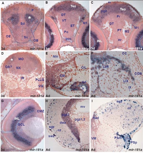
Finally, comparison of adult miR-181a and miR-181b expression reveals similar regional expression but differences at the cellular level within some areas. Good examples are the caudal hypothalamus and the facial lobe. Both miRNAs are expressed in these areas but likely in different cells. In other areas, like pretectum and tectum, telencephalon and olfactory bulb, they are likely expressed in the same cell types. A. transverse section through the larval telencephalon showing miR-181a expressing cells throughout the ventral (Sv) and dorsal (Sd) subpallium, pallium (P) and olfactory bulb (OB). The asterisk marks the strongly expressing miR-181a cell group in the central pallium, close to OB and Sd.
B. transverse section through the larval diencephalon and optic tectum (TeO) showing strongly miR-181a expressing cells in the TeO and migrated pretectal area (M1) and weakly in the preoptic area (Po), ventral thalamus (VT), dorsal thalamus (DT) and eminentia thalami (ET).
C. transverse section through the larval diencephalon and optic tectum (TeO) showing strongly miR-181a expressing cells in the TeO and migrated pretectal area (M1) and weakly in the rostral hypothalamus (Hr), periventricular (PT) and migrated (M2) posterior tuberculum, dorsal thalamus (DT) and periventricular pretectum (Pr).
D. transverse section through the larval hindbrain at the level of the posterior lateral line (PLLG) and vagal (VG) ganglia showing miR-181a expressing cells in the medulla oblongata (MO). The asterisk marks the strongly miR-181a expressing cells in the lateral medulla oblongata likely in proximity to the octaval area (OA?).
E. transverse section through the adult hypothalamus showing miR-181a expressing cells in the caudal part of the dorsal zone of the periventricular hypothalamus (Hd) around the ventricular recess (lr) and mammillary body (CM).
F. transverse section through the adult caudal hindbrain showing miR-181a expressing cells in the facial (LVII) and glossopharyngeal (LIX) lobes.
G. transverse section through the larval retina showing miR-181a expressing cells in the inner part (amacrine cells) of the inner nuclear layer (INL) and the ganglion cell layer (GCL).
H. transverse section through the adult optic tectum showing miR-181a expressing cells in the superficial gray zone (sgz), central zone (cz) and the third division of the periventricular gray zone (pgz3).
I. transverse section through the adult dorsal diencephalon and rostral optic tectum showing miR-181a expressing cells in the superficial gray zone (sgz), central zone (cz), parvocellular (PSp) and magnocellular (PSm) superficial pretectal nuclei.
|
