FIGURE

Fig. S15

ID
ZDB-FIG-080912-6
Publication
Kapsimali et al., 2007 - MicroRNAs show a wide diversity of expression profiles in the developing and mature central nervous system
Other Figures
All Figure Page
Back to All Figure Page
Fig. S15

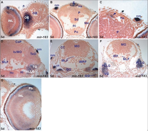
miR-183 expression in the zebrafish brain.
miR-183 expression is cell type specific in larvae. It is expressed in retinal photoreceptors and weakly in some inner nuclear layer cells, pineal cells that are again likely to be photoreceptors and perhaps also in parapineal photoreceptors. Outside of the CNS, miR-183 is expressed in cells that include peripheral sensory neuromasts, olfactory sensory neurons and hair cells of the ear. miR-182 and miR-96 show almost identical expression patterns to miR-183 (Table E) although expression is not as robust.

A. transverse section through the larval rostral brain and retina showing miR-183 expressing cells in the olfactory epithelium (OE), retinal photoreceptor (Ph) and inner nuclear (INL) layers.
B. transverse section through the larval telencephalon and rostral epithalamus showing miR-183 expressing cells in the epiphysis (E) and neuromasts (nm).
C. transverse section through the larval epithalamus and pallium (P) showing miR-183 expressing cells medial to the habenula (Ha) likely corresponding to parapineal organ (arrowhead) and neuromasts (nm).
D. transverse section through the larval midbrain and isthmus showing miR-183 expressing cells in the trigeminal (TG), anterior lateral line (ALLG) ganglia and neuromasts (nm).
E. transverse section through the larval hindbrain showing miR-183 expressing cells in the otic capsule (ot), octaval (OG) and glossopharyngeal (GG) ganglia and neuromasts (nm).
F. transverse section through the larval caudal hindbrain showing miR-183 expressing cells in the posterior lateral line (PLLG) and vagal (VG) ganglia.
G. transverse section through the larval retina showing miR-183 expressing cells in the retinal photoreceptor (Ph) and inner cellular layer (INL).

Expression Data
Gene:
Fish:
Anatomical Terms:
Stage: Day 5

Expression Detail
Antibody Labeling
Phenotype Data

Phenotype Detail
Acknowledgments
This image is the copyrighted work of the attributed author or publisher, and ZFIN has permission only to display this image to its users. Additional permissions should be obtained from the applicable author or publisher of the image.
Open Access.
Full text @ Genome Biol.