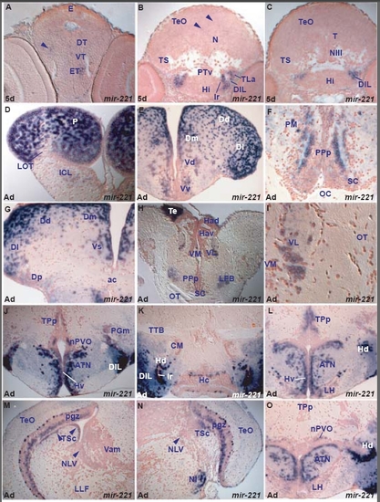
miR-221 expression in the zebrafish brain.
miR-221 shows spatially localized expression. It is strongly expressed in lateral hypothalamic areas (cells around the lateral recess, in the lateral torus and diffuse inferior lobe) and at high levels in caudal pallium and eminentia thalami. It is weakly expressed in a few rostral ventral telencephalic, thalamic, periventricular posterior tubercular, semicircular torus and rostral tectal cells (Table D). miR-221 expression is largely conserved in the adult telencephalic, hypothalamic, thalamic, posterior tubercular and semicircular torus nuclei (table I). However, there are some differences between the larval and adult miR-221 brain expression. miR-221 is expressed in adult preoptic area cells, isthmic nucleus, facial and vagal lobes, areas devoid of larval expression. miR-221 is also expressed in two cell populations in the adult tectal periventricular gray zone, area with few weakly stained cells in the larva.
miR-221 belongs to the same cluster as miR-222 and seems to largely share expression patterns but they also have subtle differences in transcript localisation. For instance, only miR-222 is expressed in the larval ventral intermediate hypothalamus, whereas only miR-221 is expressed in adult thalamic and pretectal nuclei, many adult migrated posterior tuberculum nuclei, larval and adult semicircular torus cells (tables D, I). A. transverse section through the larval diencephalon showing weakly miR-221 expressing cells in the eminentia thalami (ET) and thalamic area (arrowhead, dorsal thalamus-DT, ventral thalamus-VT).
B. transverse section through the larval diencephalon and midbrain showing miR-221 strongly expressing cells at the level of the intermediate hypothalamus (Hi) in the diffuse nucleus of the hypothalamic inferior lobe (DIL) and the lateral torus (TLa) and weakly expressing cells in the optic tectum (arrowheads, TeO).
C. transverse section through the larval hypothalamus and midbrain showing miR-221 strongly expressing cells at the level of the intermediate hypothalamus (Hi) in the diffuse nucleus of the hypothalamic inferior lobe (DIL).
D. transverse section through the adult rostral telencephalon showing robust expression of miR-221 in the dorsal telencephalon/pallium (P) and weakly in the internal cellular layer of the olfactory bulb (ICL).
E. transverse section through the adult telencephalon showing miR-221 expressing cells in the ventral (Vv) and dorsal (Vd) nuclei of the ventral telencephalon (subpallium), medial (Dm), central (Dc), dorsal (Dd) and lateral (Dl) zones of the dorsal telencephalon.
F. transverse section through the adult preoptic area at the level of the optic chiasma (posterior to section E) showing miR-221 expressing cells in the suprachiasmatic nucleus (SC), preoptic posterior parvocellular (PPp) and magnocellular (PM) nuclei.
G. transverse section through the adult telencephalon at the level of anterior commissure (ac) showing miR-221 expressing cells in the supracommissural nucleus of the ventral telencephalon (Vs), medial (Dm), dorsal (Dd), lateral (Dl) and posterior (Dp) zones of the dorsal telencephalon.
H. transverse section through the adult diencephalon showing miR-221 expressing cells in the ventrolateral thalamic nucleus (VL) and the caudal telencephalon (Te).
I. Higher magnification of the section E at the level of the thalamus showing miR-221 expressing cells in the ventrolateral thalamic nucleus (VL).
J. transverse section through the adult hypothalamus showing miR-221 expressing cells in the ventral zone of periventricular hypothalamus (Hv), anterior tuberal nucleus (ATN), diffuse nucleus of the hypothalamic inferior lobe (DIL), nucleus of the paraventricular organ (nPVO), nucleus of periventricular posterior tuberculum (TPp) and medial preglomeral nucleus (PGm).
K. transverse section through the adult hypothalamus showing miR-221 expressing cells in the caudal zone of the periventricular hypothalamus (Hc), dorsal zone of the periventricular hypothalamus (Hd, at the level of lateral ventricular recess-lr) and diffuse nucleus of the hypothalamic inferior lobe (DIL).
L. transverse section through the adult hypothalamus showing miR-221 expressing cells in the ventral (Hv) and dorsal (Hd) zones of periventricular hypothalamus, anterior tuberal nucleus (ATN), lateral hypothalamic nucleus (LH), and nucleus of periventricular posterior tuberculum (TPp).
M. transverse section through the adult isthmus and caudal midbrain showing miR-221 strongly expressing cells in the tectal periventricular gray zone (pgz), central nucleus of semicircular torus (TSc, arrowheads), and weakly in the nucleus of lateral valvula (NLV, arrowhead).
N. transverse section through the adult isthmus and caudal midbrain showing miR-221 strongly expressing cells in the tectal periventricular gray zone (pgz) of the optic tectum (TeO), central nucleus of semicircular torus (TSc), isthmic nucleus (NI) and weakly in the nucleus of lateral valvula (NLV, arrowhead).
O. transverse section through the adult hypothalamus showing miR-221 expressing cells in the anterior tuberal nucleus (ATN), lateral hypothalamic nucleus (LH) and nucleus of the paraventricular organ (nPVO).
|
