|
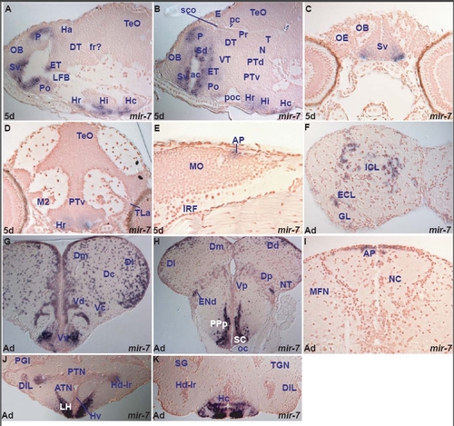
miR-7 expression in the zebrafish brain. miR-7 shows spatially localized expression, largely conserved throughout life. Larval expression is restricted to the forebrain in telencephalic, preoptic, few thalamic, eminentia thalami and ventral hypothalamic cells. In addition, it is expressed in a few cells of the area postrema at the dorsal junction of hindbrain-spinal cord (Table D). miR-7 expression is mainly conserved in telencephalic, hypothalamic nuclei and area postrema between the larval and adult zebrafish brain. However, in the adult, we observe expression in the external and internal cellular layers of the olfactory bulb. Furthermore, in contrast to the larval, the adult thalamus is devoid of miR-7 expression (Table D, I). miR-7 often shares expression in the forebrain with miR-222 but there are also some differences (table D, I). A. parasagittal section through the larval fore- and midbrain showing miR-7 expressing cells in the hypothalamus (caudal, Hc, intermediate, Hi and rostral, Hr), preoptic area (Po), eminentia thalami (ET), ventral subpallium (Sv) around the anterior commissure and pallium (P).
B. sagittal section through the larval fore- and midbrain showing miR-7 expressing cells in the hypothalamus (caudal, Hc, intermediate, Hi and rostral, Hr), preoptic area (Po), eminentia thalami (ET), ventral (Sv) and dorsal (Sd) subpallium and pallium (P).
C. transverse section through the rostral larval telencephalon showing miR-7 expressing cells in the ventral subpallium (Sv).
D. transverse section through the larval hypothalamus and midbrain showing miR-7 expressing cells in the rostral ventral hypothalamus (Hr).
E. sagittal section through the larval hindbrain showing miR-7 expressing cells in the caudal medulla oblongata (MO) at the level of the area postrema (AP).
F. transverse section through the adult olfactory bulb showing miR-7 expressing cells in the internal (ICL) and external (ECL) cellular olfactory layers.
G. transverse section through the adult telencephalon showing miR-7 expressing cells in the ventral (Vv), central (Vc) and dorsal (Vd) nuclei of the ventral telencephalon (subpallium), medial (Dm), central (Dc) and lateral (Dl) zones of the dorsal telencephalon (pallium). Expression is largely absent from cells lining the ventricles.
H. transverse section through the caudal adult telencephalon and rostral diencephalon (at the level of the optic chiasma, oc) showing miR-7 expressing cells in the suprachiasmatic nucleus (SC), posterior parvocellular preoptic nucleus (PPp), dorsal endopeduncular nucleus (ENd, part of the eminentia thalami), posterior nucleus of the ventral telencephalon (Vp), posterior (Dp), medial (Dm), dorsal (Dd) and lateral (Dl) zones of the dorsal telencephalon.
I. transverse section through the caudal medulla oblongata (MO) showing miR-7 expressing cells in the area postrema (AP).
J. transverse section through the adult hypothalamus showing miR-7 expressing cells in the lateral hypothalamic nucleus (LH), ventral zone of the periventricular hypothalamus (Hv), dorsal zone of the periventricular hypothalamus at the level of the lateral hypothalamic ventricular recess (Hd-lr) and diffuse nucleus of the inferior hypothalamic lobe (DIL).
K. transverse section through the adult hypothalamus (caudal to section J) showing miR-7 expressing cells in the caudal zone of the periventricular hypothalamus (Hc).
|