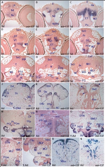
miR-137 expression in the zebrafish brain.
miR-137 shows spatially localized, conserved expression in specific larval and adult brain nuclei or cells (Table C, H). miR-137 expression is conserved in many larval and adult areas including the subpallium, ventro-medial and caudal pallium, preoptic area, dorsal thalamus, hypothalamus and ventral posterior tubercular area. Given the good correspondence of localized expression between larval and adult brains, this allows us to annotate specific nuclei in the larval tegmentum, isthmus and medulla oblongata. We suggest that the cells in the larval tegmentum and medulla oblongata correspond to the midbrain dorsal tegmental nucleus, lateral nucleus of cerebellar valvula, isthmic nucleus, facial and glossopharyngeal/vagal lobes, vagal motor nucleus and area postrema. Despite the well-conserved pattern of expression between larval and adult zebrafish brain, we observe minor differences. miR-137 is expressed in adult dorsal lateral habenular cells and a few tectal periventricular gray zone and migrated posterior tuberculum cells. A. transverse section through the larval telencephalon showing miR-137 expressing cells in the ventral (Sv) and dorsal (Sd) subpallium and pallium (P).
B. transverse section through the caudal telencephalon and epithalamus showing miR-137 expressing cells in the ventral subpallium(Sv)/preoptic area (Po), dorsal subpallium (Sd), migrated telencephalic area (M4) and pallium (P).
C. transverse section through the caudal telencephalon and epithalamus showing miR-137 expressing cells in the pallium (P) and eminentia thalami (ET).
D. transverse section through the larval diencephalon and rostral optic tectum showing miR-137 expressing cells in the eminentia thalami (ET), ventral thalamus (VT) and dorsal thalamus (DT).
E. transverse section through the larval diencephalon and rostral optic tectum (caudal to section D) showing miR-137 expressing cells in the eminentia thalami (ET), ventral thalamus (VT) and dorsal thalamus (DT).
F. transverse section through the larval diencephalon and midbrain showing miR-137 expressing cells in the rostral hypothalamus (Hr), dorsal periventricular posterior tuberculum (PTd) and dorsal thalamus (DT).
G. transverse section through the larval diencephalon and midbrain showing miR-137 expressing cells in the intermediate hypothalamus (Hi), lateral hypothalamic torus (TLa) and ventral periventricular posterior tuberculum (PTv).
H. transverse section through the larval diencephalon and midbrain showing miR-137 expressing cells in the lateral hypothalamic torus (TLa), ventral periventricular posterior tuberculum (PTv) and midbrain dorsal tegmental nucleus (DTN).
I. transverse section through the larval caudal midbrain and rostral hindbrain at the level of the facial ganglion (FG) showing miR-137 expressing cells in the isthmic area (Is) including the isthmic nucleus (NI).
J. transverse section through the adult telencephalon at the level of the anterior commissure (ac) showing miR-137 expressing cells in the supracommissural nucleus of the ventral (subpallial) telencephalic area (Vs), posterior (Dp), lateral (Dl), dorsal (Dd), medial (Dm) zones of the dorsal (pallial) telencephalic area and the dorsal entopeduncular nucleus (ENd).
K. transverse section through the adult caudal telencephalon at the level of the optic chiasma (oc) showing miR-137 expressing cells in the posterior (Dp), lateral (Dl) and medial (Dm) zones of the dorsal (pallial) telencephalic area, posterior preoptic parvocellular (PPp) and suprachiasmatic (SC) nuclei.
L. transverse section through the adult epithalamus showing miR-137 expressing cells in the dorsal habenular nucleus (Had, arrowheads).
M. transverse section through the adult diencephalon showing miR-137 expressing cells in the ventral zone of the hypothalamus (Hv), anterior tuberal nucleus (ATN), dorsal zone of the periventricular hypothalamus (Hd), lateral torus (TLa), posterior tuberal nucleus (PTN), medial preglomeral nucleus (PGm), posterior thalamic nucleus (Pt) and central posterior thalamic nucleus (CP). For higher magnification of the thalamus, see section N.
N. Higher magnification of a transverse section (level caudal to section M) through the adult dorsal diencephalon, at the level of the posterior commissure (pc) showing miR-137 expressing cells in the central posterior thalamic nucleus (CP).
O. transverse section through the young adult diencephalon and midbrain showing miR-137 expressing cells in the ventral zone of the hypothalamus (Hv), lateral hypothalamic nucleus (LH), dorsal zone of the periventricular hypothalamus around the lateral hypothalamic ventricular recess (Hd-lr), diffuse nucleus of inferior lobe (DIL), lateral torus (TLa), posterior tuberal nucleus (PTN), ventral part of the nucleus of the posterior periventricular tuberculum (TPp), posterior thalamic nucleus/lateral preglomeral nucleus (Pt/PGl) and midbrain dorsal tegmental nucleus (DTN).
P. Higher magnification of the dorsal part of the transverse section P showing miR-137 expressing cells in the midbrain dorsal tegmental nucleus (DTN), ventral part of the nucleus of the posterior periventricular tuberculum (TPp) and a few cells in the proximity of the mesencephalic ventricle (arrowhead).
Q. transverse section through the young adult caudal hypothalamus, midbrain and isthmus showing miR-137 expressing cells in the caudal zone of the hypothalamus (Hc), diffuse nucleus of the hypothalamic inferior lobe (DIL), isthmic nucleus (NI), nucleus of lateral cerebellar valvula (NLV), central nucleus of semicircular torus (TSc) and optic tectum (TeO).
R. transverse section through the adult caudal hindbrain at the level of the vagal nerve showing miR-137 expressing cells in the vagal lobe (LX) and vagal motor nucleus (NXm).
S. transverse section through the adult hindbrain at the junction with the spinal cord (caudal to section R) showing miR-137 expressing cells in the area postrema (AP) and commissural nucleus of Cajal (NC).
|
