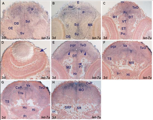
let-7a expression in the zebrafish brain.
let-7a, let-7b and let-7c are expressed in both proliferating and differentiating cells. let-7b and let-7c differ in their sequence in only one nucleotide located outside the seed region. They share similar regional expression in the larval brain with two differences: let-7b is expressed in the retinal ciliary marginal zone and pineal cells whereas let-7a and let-7c are absent (table A). let-7a, let-7b and let-7c mainly conserve their regional expression between larval and adult brain (tables A, F).
let-7a, let-7b and let-7c are expressed in many proliferating and differentiating cells of the larval fore-, mid- and hindbrain with the exception of some areas such as hypothalamic nuclei (caudal hypothalamus, diffuse nucleus of inferior lobe, lateral torus) interpeduncular nucleus, locus coereleus, raphe and reticular formation. We detected only minor differences at the regional level between larval and adult brain expression. For example, let-7b and let-7c are expressed in some adult but not larval hypothalamic lateral torus and superior raphe cells (tables A, F). A. transverse section through the larval telencephalon showing let-7a expressing cells in the ventral (Sv) and dorsal (Sd) subpallium and pallium (P).
B. transverse section through the larval telencephalon and epithalamus showing let-7a expressing cells in the ventral (Sv) and dorsal (Sd) subpallium, pallium (P), migrated telencephalic area (M4) and habenula (Ha). Pineal cells (E) are devoid of expression.
C. transverse section through the larval diencephalon and rostral optic tectum showing let-7a expressing cells in the preoptic area (Po), eminentia thalami (ET), ventral thalamus (VT) and dorsal (DT) thalamus, periventricular (Pr) and migrated (M1) pretectum and optic tectum (TeO, including the tectal proliferative zone, m).
D. transverse section through the larval retina (dorsal to the right) devoid of let-7a expressing cells. The arrow points at the ciliary marginal zone.
E. transverse section through the larval diencephalon and midbrain showing mainly periventricular (arrowheads) let-7a expressing cells in the rostral hypothalamus (Hr), periventricular (PT) and migrated (M2) posterior tuberculum, dorsal thalamus (DT), tegmentum (T) and tectal periventricular gray zone (pgz).
F. transverse section through the larval hypothalamus, midbrain and rostral hindbrain showing let-7a expressing cells in the intermediate hypothalamus (Hi, area of the periventricular hypothalamic recess-lr), semicircular torus (TS), tegmentum (T), tectal periventricular gray zone (pgz) and cerebellar valvula (Va).
G. transverse section through the larval hypothalamus, midbrain and rostral hindbrain showing let-7a expressing cells in the intermediate (Hi) and caudal (Hc) hypothalamus, semicircular torus (TS), tegmentum/isthmic area (T/Is), periventricular gray zone (pgz) of the optic tectum (TeO), cerebellar valvula (Va) and cerebellar plate (CeP).
H. transverse section through the larval hindbrain at the level of the otic capsule (ot) showing let-7a expressing cells in the medulla oblongata (MO).
|
