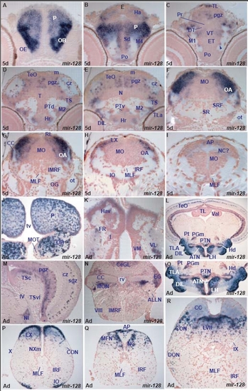
miR-128 expression in the zebrafish brain.
miR-128 shows spatially localized, conserved expression in specific larval and adult brain nuclei or cells. In addition, miR-128 expression shows major quantitative differences in levels of expression between structures in the larval brain.
At 5dpf, miR-128 is expressed in the entire olfactory bulb, pallium, and lateral medulla oblongata. In these regions, it is upregulated in the olfactory bulb, caudal pallial and lateral medulla oblongata cells. It is also expressed in distinct cells in the habenula, ventral thalamus, posterior tuberculum, lateral hypothalamus, periventricular and central tectal zones, semicircular torus and area postrema. It conserves its expression in these areas in the adult brain and given the good overall regional similarities in miR-128 expression between 5dpf and adult brain we suggest that the 5dpf expression in the medulla oblongata corresponds to cells of the presumptive octaval area, facial and vagal lobes, area postrema and commissural nucleus of Cajal. One difference in the expression pattern of miR-128 between larval and adult brain is expression in the adult inferior olive (Tables C, H). A. transverse section through the rostral larval telencephalon showing miR-128 expressing cells in the olfactory bulb (OB) and pallium (P).
B. transverse section through the caudal telencephalon and epithalamus showing miR-128 expressing cells in the preoptic area (Po), dorsal subpallium (Sd), migrated telencephalic area (M4), pallium (P) and habenula (Ha).
C. transverse section through the larval diencephalon and optic tectum showing miR-128 expressing cells in the lateral eminentia thalami (ET, arrowhead), ventral thalamus (VT), migrated pretectal area (M1), periventricular gray zone (pgz) and longitudinal torus (TL) of the optic tectum.
D. transverse section through the larval hypothalamus and midbrain showing miR-128 expressing cells in the rostral hypothalamus (Hr), migrated posterior tubercular area (M2), semicircular torus (TS), periventricular gray (pgz) and central zone (cz) of the optic tectum (TeO).
E. transverse section through the larval hypothalamus and midbrain showing miR-128 expressing cells in the rostral hypothalamus (Hr), lateral torus (TLa), ventral periventricular (PTv) and migrated posterior tubercular area (M2), semicircular torus (TS), periventricular gray (pgz) and central zone (cz) of the optic tectum (TeO).
F. transverse section through the larval hindbrain at the level of the otic capsule (ot) showing miR-128 expressing cells in the lateral medulla oblongata (MO), including the octaval area (OA).
G. transverse section through the larval hindbrain at the level of the octaval ganglion (OG) showing miR-128 expressing cells in the lateral medulla oblongata (MO), including the octaval area (OA).
H. transverse section through the larval hindbrain caudal to the posterior lateral line ganglion showing miR-128 expressing cells in the lateral medulla oblongata (MO) likely in the octaval area (OA) and vagal lobe (LX).
I. transverse section through the larval caudal medulla oblongata (MO) showing miR-128 expressing cells in the area postrema (AP) and likely the commissural nucleus of Cajal (NC).
J. transverse section through the rostral adult telencephalon showing miR-128 expressing cells in the dorsal telencephalon/pallium (P) and olfactory bulb (OB) with the exception of the area of the medial olfactory tract (MOT).
K. transverse section through the adult epithalamus and thalamus showing miR-128 in the dorsal part of the ventral habenula (Hav) and ventrolateral thalamic nucleus (VL).
L. transverse section through the adult hypothalamus and midbrain showing miR-128 expressing cells in the lateral hypothalamic nucleus (LH), anterior tuberal nucleus (ATN), dorsal zone of the periventricular hypothalamus (Hd), diffuse nucleus of the inferior hypothalamic lobe (DIL), lateral torus (TLa), posterior tuberal nucleus (PTN), medial preglomeral nucleus (PGm), posterior thalamic nucleus (Pt) and optic tectum (TeO). For higher magnifications of the hypothalamus and optic tectum, see pictures M and O.
M. transverse section through the adult midbrain and isthmus (caudal to section L) showing miR-128 expressing cells in the isthmic nucleus (NI), central nucleus of semicircular torus (TSc), periventricular gray (pgz) and superficial grey (sgz) zones of the optic tectum. The asterisk marks the proliferative tectal zone devoid of miR-128 expression.
N. transverse section through the adult hindbrain at the level of the octaval nerve (VIII) showing miR-128 expressing cells in the medial octavolateral nucleus (MON), cerebellar granular eminence (EG) and granular cerebellar layer (CeGL).
O. higher magnification of section L at the level of the adult hypothalamus showing miR-128 expressing cells in the lateral hypothalamic nucleus (LH), anterior tuberal nucleus (ATN), dorsal zone of the periventricular hypothalamus (Hd), diffuse nucleus of the inferior hypothalamic lobe (DIL), lateral torus (TLa), posterior tuberal nucleus (PTN), medial preglomeral nucleus (PGm) and posterior thalamic nucleus (Pt).
P. transverse section through the adult hindbrain at the level of the vagal nerve (X, section caudal to R) showing miR-128 expressing cells in the inferior olive (IO), caudal octavolateral nucleus (CON) and vagal lobe (LX).
Q. transverse section through the adult hindbrain at the junction with the spinal cord (section caudal to P) showing miR-128 expressing cells in the area postrema (AP), commissural nucleus of Cajal (NC) and medial funicular nucleus (MFN).
R. transverse section through the adult hindbrain at the level of the cerebellar crest (CC, section caudal to N) showing miR-128 expressing cells in the descending octaval nucleus (DON), caudal octavolateral nucleus (CON) and facial lobe (LVII).
|
