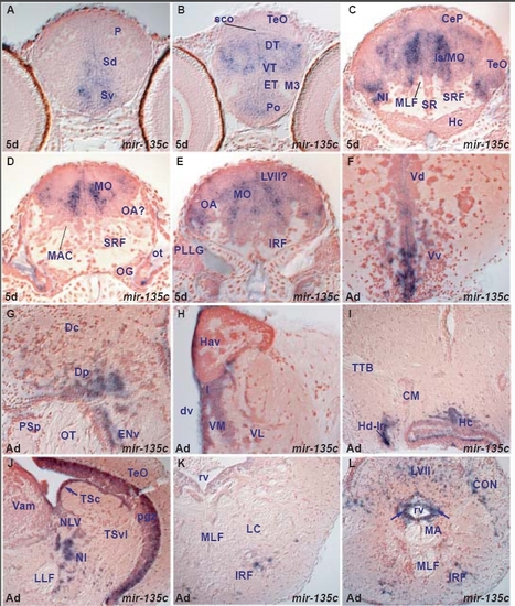
miR-135c expression in the zebrafish brain.
miR-135c is expressed in both periventricular and differentiating cells with a more restricted pattern compared to miR-9. miR-135c expression is mainly conserved between larval and adult brain but also shows some differences (Tables B,G). miR-135c expression is conserved in both larval and adult subpallium, caudal pallium, habenular, preoptic, thalamic, hypothalamic, pretectal, tegmental, cerebellar, isthmic and octaval nuclei. It is expressed in additional adult areas, devoid of larval expression, such as the caudal hypothalamus, periventricular and migrated posterior tuberculum nuclei, inferior raphe and reticular formation. In other hindbrain areas, it is difficult to draw conclusion about the conservation of miR-135c expression from larval to adult stages: miR-135c is robustly expressed in medial and central columns throughout the larval isthmic area and medulla oblongata but this is not the case in the adult. miR-135c is expressed in many cells of the adult facial and vagal lobes, and this expression may correspond to part of the larval one. A. transverse section through the larval telencephalon showing miR-135c expressing cells in the ventral (Sv) and dorsal (Sd) subpallium.
B. transverse section through the larval diencephalon and rostral optic tectum showing miR-135c expressing cells in the preoptic area (Po), eminentia thalami (ET), ventral (VT) and dorsal thalamus (DT).
C. transverse section through the larval caudal midbrain, hypothalamus, cerebellum and isthmus/medulla oblongata showing miR-135c expressing cells in the isthmic nucleus (NI), cerebellar plate (CeP), optic tectum (TeO) and isthmic/medulla oblongata areas (Is/MO).
D. transverse section through the larval hindbrain at the level of the octaval ganglion (OG) showing miR-135c expressing cells in the medulla oblongata (MO).
E. transverse section through the larval hindbrain at the level of the posterior lateral line ganglion (PLLG) showing miR-135c expressing cells in the medulla oblongata (MO) likely including the presumptive octaval area (OA).
F. transverse section through the adult telencephalon showing miR-135c expressing cells lining or lateral to the telencephalic ventricle in the subpallium (ventral nucleus of the ventral telencephalon,Vv, dorsal nucleus of the ventral telencephalon,Vd).
G. transverse section through the adult caudal telencephalon showing miR-135c expressing cells in the posterior zone of the dorsal telencephalon (Dp) and ventral part of the entopeduncular nucleus (ENv).
H. transverse section through the adult dorsal diencephalon showing miR-135c expressing cells lining or close to the diencephalic ventricle (dv) in the ventral habenular nucleus (Hav), intermediate (I), ventromedial (VM) and ventrolalateral (VL) thalamic nuclei.
I. transverse section through the adult midbrain and hypothalamus showing miR-135c expressing cells lining/close to the ventricle in the dorsal (Hd-lr, around the lateral ventricular recess) and caudal (Hc) zones of the periventricular hypothalamus.
J. transverse section through the adult caudal midbrain and isthmus showing miR-135c expressing cells in the tectal periventricular gray zone (pgz), periventricular central semicircular torus (arrow, TSc) nucleus of lateral valvula (NLV) and isthmic nucleus (NI).
K. transverse section through the adult isthmus showing miR-135c expressing cells in the inferior reticular formation (IRF).
L. transverse section through the caudal medulla oblongata showing miR-135c expressing cells in the inferior reticular formation (IRF), caudal octavolateral nucleus (CON), facial lobe (LVII) and surrounding the rhombencephalic ventricle (rv, arrows).
|
