
Fig. 3
- ID
- ZDB-FIG-070418-13
- Publication
- Guner et al., 2007 - Cloning of zebrafish nkx6.2 and a comprehensive analysis of the conserved transcriptional response to Hedgehog/Gli signaling in the zebrafish neural tube
- Other Figures
- All Figure Page
- Back to All Figure Page
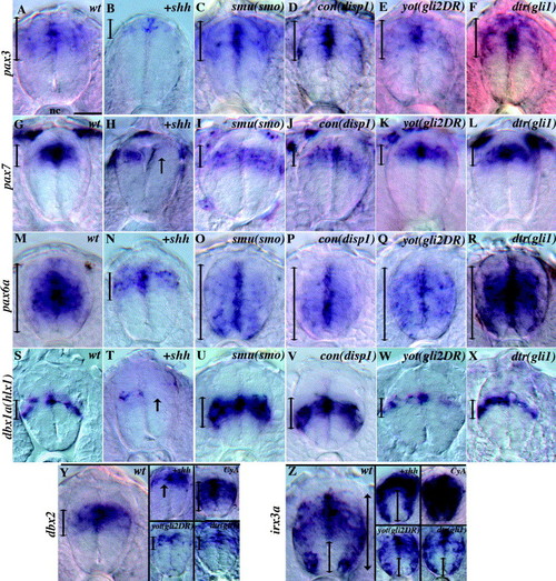
Class I gene expression in zebrafish and regulation by Hh. (A) pax3 is expressed in the dorsal-most cells of the spinal cord, including the roof plate. (B) pax3 expression is absent or restricted dorsally in shh mRNA injected embryos. (C) In smu(smo) mutants, spinal cord pax3 expression appears normal. (D–F) The expression of pax3 is unaffected in the neural tubes of con(disp1) (D), yot(gli2DR) (E), and dtr(gli1) (F) mutants. (G) pax7 is expressed in the dorsal spinal cord, but not in the roof plate. (H) pax7 expression is reduced or absent in shh mRNA-injected embryos. (I–L) The expression of pax7 is unaltered in smu(smo) (I), con(disp1) (J), yot(gli2DR) (K), and dtr(gli1) (L) mutant embryos. (M) pax6a is expressed throughout the spinal cord with the exception of the most ventral and most dorsal cells (arrow). (N) pax6a is reduced in shh mRNA-injected embryos. (O–R) pax6a expression is ventrally expanded in smu(smo) (O), con(disp1) (P), yot(gli2DR) (Q), and dtr(gli1) (R) mutants (arrows). (S) dbx1a(hlx1) is expressed in a band of cells in the middle of the D/V axis of the spinal cord. (T) dbx1a(hlx1) expression is reduced or absent after shh mRNA injection (arrow). (U–X) dbx1a(hlx1) expression is ventrally expanded in smu(smo) (U), and con(disp1) (V) mutants but is not altered in yot(gli2DR) (W), and dtr(gli1) (X) mutants. (Y) dbx2 is expressed in the mid-dorsal cells of the spinal cord. (Y) The expression of dbx2 is reduced an restricted to the dorsal most cells of the spinal cord in shh mRNA-injected embryos and dbx2 expression is expanded ventrally in cyclopamine treated embryos. dbx2 expression appears unaffected in yot(gli2DR) and dtr(gli1) mutants. (Z) irx3a is expressed in lateral and mid-dorsal cells. In shh mRNA-injected embryos, medial expression of irx3a is repressed while lateral irx3a expression remains unaffected. In cyclopamine treated embryos irx3a is expressed throughout the neural tube with the exception of the medial floor plate. The expression of irx3a appears unaffected in yot(gli2DR) and dtr(gli1) mutant embryos. All panels show spinal cord cross sections of 24 hpf embryos. Double-ended arrows show dorsal and ventral extent of gene expression. In general, labeling intensity appeared similar in wild type and mutant embryos. Scale bar: 15 μm (33 μm in Y and Z mutant panels). |
| Genes: | |
|---|---|
| Fish: | |
| Condition: | |
| Anatomical Terms: | |
| Stage: | Prim-5 |
Reprinted from Gene expression patterns : GEP, 7(5), Guner, B., and Karlstrom, R.O., Cloning of zebrafish nkx6.2 and a comprehensive analysis of the conserved transcriptional response to Hedgehog/Gli signaling in the zebrafish neural tube, 596-605, Copyright (2007) with permission from Elsevier. Full text @ Gene Expr. Patterns