
Fig. 7
- ID
- ZDB-FIG-050311-4
- Publication
- Yimlamai et al., 2005 - The zebrafish down syndrome cell adhesion molecule is involved in cell movement during embryogenesis
- Other Figures
- All Figure Page
- Back to All Figure Page
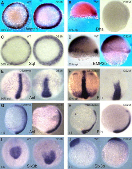
Marker gene in situ of dscam knockdown embryos. (A and C) Animal pole view. (B and D) Lateral view, dorsal is right, animal pole is up. (E and F) Dorsal view, tailbud is at the bottom. (G, H, and J) Lateral view, tailbud is at the bottom. (I) Rostral view. (A and C) Wnt11 and Squint/Nodal at 30% epiboly, respectively. (B and D) Dharma and Bmp2b at 50% epiboly, respectively. (E and F) Axial and floating head at 90% epiboly, respectively. (E and F) Axial and floating head at 1 somite, respectively. (I and J) Six3b at 8 somites. All controls show representative samples. DS2M injections show severe phenotype in at least five embryos in samples of twenty. |
| Genes: | |
|---|---|
| Fish: | |
| Condition: | |
| Knockdown Reagent: | |
| Anatomical Term: | |
| Stage Range: | 30%-epiboly to 5-9 somites |
Reprinted from Developmental Biology, 279(1), Yimlamai, D., Konnikova, L., Moss, L.G., and Jay, D.G., The zebrafish down syndrome cell adhesion molecule is involved in cell movement during embryogenesis, 44-57, Copyright (2005) with permission from Elsevier. Full text @ Dev. Biol.