
FIGURE
Fig. 6
- ID
- ZDB-FIG-071004-14
- Publication
- Yimlamai et al., 2005 - The zebrafish down syndrome cell adhesion molecule is involved in cell movement during embryogenesis
- Other Figures
- All Figure Page
- Back to All Figure Page
Fig. 6
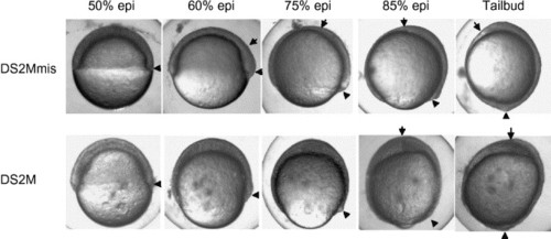
Time lapse images of control and dscam morphants during gastrulation. Images are of a single embryo photographed at the indicated stages. Arrowhead represents the tailbud. Arrow represents leading edge of the polster. |
Expression Data
Expression Detail
Antibody Labeling
Phenotype Data
Phenotype Detail
Acknowledgments
This image is the copyrighted work of the attributed author or publisher, and
ZFIN has permission only to display this image to its users.
Additional permissions should be obtained from the applicable author or publisher of the image.
Reprinted from Developmental Biology, 279(1), Yimlamai, D., Konnikova, L., Moss, L.G., and Jay, D.G., The zebrafish down syndrome cell adhesion molecule is involved in cell movement during embryogenesis, 44-57, Copyright (2005) with permission from Elsevier. Full text @ Dev. Biol.