- Title
-
Identification of tissues and patterning events required for distinct steps in early migration of zebrafish primordial germ cells
- Authors
- Weidinger, G., Wolke, U., Köprunner, M., Klinger, M., and Raz, E.
- Source
- Full text @ Development
|
Migration of PGCs in wild-type embryos. The PGCs are labeled using the vasa mRNA probe (dark blue) and other structures are labeled in red or in light blue with the probes indicated. Embryos at somitogenesis stages were deyolked and flattened. (A-C) The three basic initial PGC arrangements relative to the dorsal aspect of the embryo are shown at the dome stage (upper panel), where dorsal is marked by the chordino expression domain. At 60% epiboly (middle panel) and the 2-somite stage (lower panel) also three classes of arrangements are found at frequencies expected from the three initial arrangements at the dome stage. The 60% epiboly stages are stained with chordino (PGC clusters are marked with arrowheads), the 2-somite stages with myoD marking the adaxial cells, papc expressed in the segmental plate and the forming somites, and pax8 staining the otic placodes. Dome and 60% epiboly stages are shown in animal view with dorsal up. (D,E) PGCs align along the border of the trunk mesoderm that expresses papc both on the dorsal (D) and the ventral (E) side at the 90% epiboly stage. Note that no PGCs are found on the notochord, which is devoid of papc staining in D. (F) A 1-somitestage embryo showing the alignment of posterior PGCs (arrowheads) at the lateral border of the broad expression domain of pax8 in the pronephric anlage. Pax8 also stains the otic placodes and myoD the adaxial cells (dark blue). (G,H) Cross-sections of 4-somite-stage embryos after whole-mount in situ hybridization with vasa. (G) At the level of somite 1, two medially located PGCs are seen in contact with the YSL and the overlying paraxial mesoderm (arrowheads), whereas two laterally located PGCs have lost contact with the YSL and extend up to the ectoderm (arrows). (H) A posterior trailing PGC (arrow) at the level of somite 7 found at the lateral margin of the mesoderm in contact with the YSL and the ectoderm. (I) 8-somite-stage embryo stained with myoD (adaxial cells and somites) and pax2.1 (pronephros, otic placodes, midbrain-hindbrain boundary and eye anlagen). There is one ectopic anterior PGC present in this wild-type embryo (arrow). (J) A 16- somite-stage embryo stained with myoD. Note that the PGC clusters have shifted towards the posterior, whereas trailing cells have migrated anteriorly. (K,L) PGCs are located in two lateral lines at the anterior end of the yolk extension at 24 hpf as seen in lateral (K) and dorsal (L) view. |
|
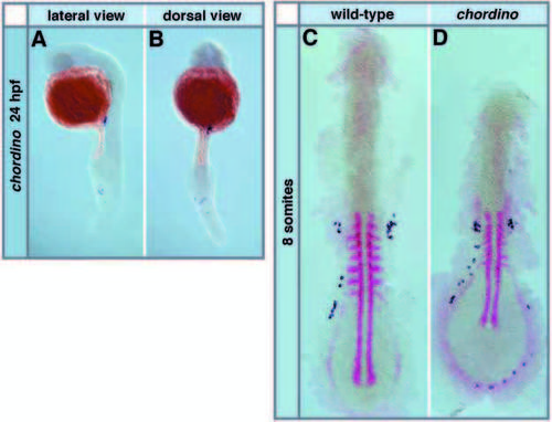
Migration of PGCs in ventralized chordino mutant embryos. The PGCs are labeled using the vasa probe (dark blue) and other structures are labeled in red with the probes indicated. (A,B) Ectopic PGCs are found around the expanded blood forming region in the tail of chordino mutants at 24 hpf; (A) lateral view, (B) dorsal view. (C,D) At the 8- somite stage (stained with myoD and gata2 labeling the Intermediate Cell Mass), more PGCs are found in the expanded ventral-posterior positions in chordino mutants (D) than in wild-type siblings (C) (see also Table 1). EXPRESSION / LABELING:
PHENOTYPE:
|
|
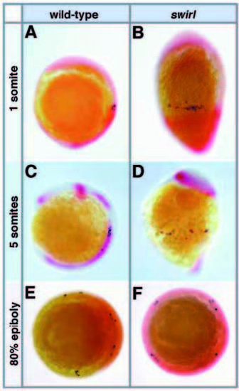
The PGCs are found in random dorsoventral positions in dorsalized swirl mutants, but all are located at the same anteroposterior level. All embryos were stained with vasa in blue and a second staining in red was done with the probes indicated. (A,B) At the 1-somite stage, an alignment of PGCs anterior to trunk mesoderm (labeled by papc and myoD) is observed in lateral view of wild-type (A) and swirl mutant (B) embryos (anterior up, dorsal right), but PGCs are found all around the dorsoventral axis in swirl mutant embryos. (C,D) Lateral view of 5-somite-stage embryos stained with pax2.1 and myoD (here only the adaxial cells are labeled by this probe). PGCs are still dispersed around the circumference of the embryo in swirl mutants (D); they do not cluster together as they do in wild-type embryos at this stage (C) (see Fig. 1I for a dorsal view of wild-type). (E,F) PGCs have converged towards the dorsal in wildtype (E), but not swirl mutant (F) embryos at 80% epiboly. Embryos are shown in vegetal view with the dorsal marked by chordino expression in the wild-type (E) and circumferential chordino expression in the swirl mutant (F). EXPRESSION / LABELING:
PHENOTYPE:
|
|
PGC migration phenotype of knypek and floating head mutant embryos. All embryos were stained with vasa in dark blue and other probes in red or light blue as indicated. (A) Trailing PGCs align along the abnormally located pronephros (stained with pax2.1, adaxial cells and somites with myoD) in knypek mutant embryos at the 7- somite stage. (B,C) Normal arrangement of PGCs in wild-type (B) and floating head mutant (C) embryos at the 8-somite stage stained with myoD and pax2.1. (D,E) Dorsal view of embryos at the 90% epiboly stage showing that dorsally located PGCs do not migrate away from the dorsal midline in floating head mutant embryos. (D) In wild-type embryos, PGCs are not found at the region of the forming notochord marked by lack of papc staining, but they extend into the dorsal midline that expresses papc in floating head mutant embryos (E). EXPRESSION / LABELING:
|
|
Ectopic PGCs are found in spadetail mutant embryos. All embryos were stained with vasa in blue and other probes in red as indicated. (A,B) Ectopic anterior PGCs are seen between the midbrain-hindbrain boundary and the otic vesicle in 24 hpf stage spadetail mutant embryos and ectopic posterior PGCs are frequently observed in the tail; (A) lateral view, (B) dorsal view. (C,D) PGC alignment at the anterior border of the papc expresssion domain at the 80% epiboly stage as seen in a dorsal view of a wildtype embryo (C) is lost in spadetail mutants (D). (E,F) At the 3- somite stage, alignment of PGCs at the level of the 1st somite as seen in wildtype embryos (E) is lost in spadetail mutants (F) stained with myoD, papc and pax8. (G,H) At the 6- somite stage (embryos stained with myoD and pax2.1), the main clusters of PGCs located at the 1st to the 3rd somite in wild-type embryos (G) are found closer to the otic placodes in spadetail mutants (H) and ectopic anterior PGCs are found mainly in between the midbrainhindbrain boundary and the otic placodes in the mutants. EXPRESSION / LABELING:
PHENOTYPE:
|
|
PGCs maintain their fate at ectopic locations. All embryos were stained with the vasa probe in blue. (A, B) Double staining of wild-type (A) and spadetail mutant (B) embryos with vasa and hoxa2 both in blue shows that anterior ectopic PGCs (arrows) are located at the anteroposterior level of the 2nd branchial arch (brackets) at 24 hpf. (C, D) Ectopic vasa-expressing cells maintain PGC morphology. Cross-section of a spadetail mutant (C) and a wild-type (D) embryo at the indicated levels. Note that both the ectopic vasa-positive cells in C and the PGCs located in the correct position in D are large and show a distinct nuclear shape (see insert). |


