
FIGURE
Fig. 4
- ID
- ZDB-FIG-140319-44
- Publication
- Weidinger et al., 1999 - Identification of tissues and patterning events required for distinct steps in early migration of zebrafish primordial germ cells
- Other Figures
- All Figure Page
- Back to All Figure Page
Fig. 4
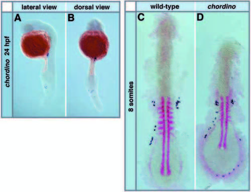
Migration of PGCs in ventralized chordino mutant embryos. The PGCs are labeled using the vasa probe (dark blue) and other structures are labeled in red with the probes indicated. (A,B) Ectopic PGCs are found around the expanded blood forming region in the tail of chordino mutants at 24 hpf; (A) lateral view, (B) dorsal view. (C,D) At the 8- somite stage (stained with myoD and gata2 labeling the Intermediate Cell Mass), more PGCs are found in the expanded ventral-posterior positions in chordino mutants (D) than in wild-type siblings (C) (see also Table 1). |
Expression Data
| Genes: | |
|---|---|
| Fish: | |
| Anatomical Terms: | |
| Stage Range: | 5-9 somites to Prim-5 |
Expression Detail
Antibody Labeling
Phenotype Data
| Fish: | |
|---|---|
| Observed In: | |
| Stage Range: | 5-9 somites to Prim-5 |
Phenotype Detail
Acknowledgments
This image is the copyrighted work of the attributed author or publisher, and
ZFIN has permission only to display this image to its users.
Additional permissions should be obtained from the applicable author or publisher of the image.
Full text @ Development