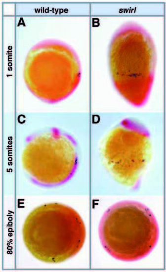
The PGCs are found in random dorsoventral positions in dorsalized swirl mutants, but all are located at the same anteroposterior level. All embryos were stained with vasa in blue and a second staining in red was done with the probes indicated. (A,B) At the 1-somite stage, an alignment of PGCs anterior to trunk mesoderm (labeled by papc and myoD) is observed in lateral view of wild-type (A) and swirl mutant (B) embryos (anterior up, dorsal right), but PGCs are found all around the dorsoventral axis in swirl mutant embryos. (C,D) Lateral view of 5-somite-stage embryos stained with pax2.1 and myoD (here only the adaxial cells are labeled by this probe). PGCs are still dispersed around the circumference of the embryo in swirl mutants (D); they do not cluster together as they do in wild-type embryos at this stage (C) (see Fig. 1I for a dorsal view of wild-type). (E,F) PGCs have converged towards the dorsal in wildtype (E), but not swirl mutant (F) embryos at 80% epiboly. Embryos are shown in vegetal view with the dorsal marked by chordino expression in the wild-type (E) and circumferential chordino expression in the swirl mutant (F).
|
