Fig. 1
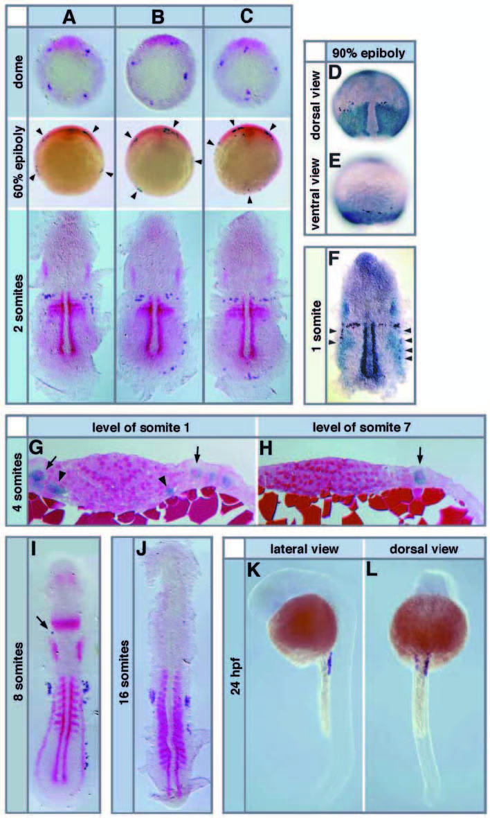
Fig. 1 Migration of PGCs in wild-type embryos. The PGCs are labeled using the vasa mRNA probe (dark blue) and other structures are labeled in red or in light blue with the probes indicated. Embryos at somitogenesis stages were deyolked and flattened. (A-C) The three basic initial PGC arrangements relative to the dorsal aspect of the embryo are shown at the dome stage (upper panel), where dorsal is marked by the chordino expression domain. At 60% epiboly (middle panel) and the 2-somite stage (lower panel) also three classes of arrangements are found at frequencies expected from the three initial arrangements at the dome stage. The 60% epiboly stages are stained with chordino (PGC clusters are marked with arrowheads), the 2-somite stages with myoD marking the adaxial cells, papc expressed in the segmental plate and the forming somites, and pax8 staining the otic placodes. Dome and 60% epiboly stages are shown in animal view with dorsal up. (D,E) PGCs align along the border of the trunk mesoderm that expresses papc both on the dorsal (D) and the ventral (E) side at the 90% epiboly stage. Note that no PGCs are found on the notochord, which is devoid of papc staining in D. (F) A 1-somitestage embryo showing the alignment of posterior PGCs (arrowheads) at the lateral border of the broad expression domain of pax8 in the pronephric anlage. Pax8 also stains the otic placodes and myoD the adaxial cells (dark blue). (G,H) Cross-sections of 4-somite-stage embryos after whole-mount in situ hybridization with vasa. (G) At the level of somite 1, two medially located PGCs are seen in contact with the YSL and the overlying paraxial mesoderm (arrowheads), whereas two laterally located PGCs have lost contact with the YSL and extend up to the ectoderm (arrows). (H) A posterior trailing PGC (arrow) at the level of somite 7 found at the lateral margin of the mesoderm in contact with the YSL and the ectoderm. (I) 8-somite-stage embryo stained with myoD (adaxial cells and somites) and pax2.1 (pronephros, otic placodes, midbrain-hindbrain boundary and eye anlagen). There is one ectopic anterior PGC present in this wild-type embryo (arrow). (J) A 16- somite-stage embryo stained with myoD. Note that the PGC clusters have shifted towards the posterior, whereas trailing cells have migrated anteriorly. (K,L) PGCs are located in two lateral lines at the anterior end of the yolk extension at 24 hpf as seen in lateral (K) and dorsal (L) view.

