
- Title
-
Protective Effect of myo-Inositol Against Decitabine-Induced Neural Tube Defects in Embryonic Zebrafish
- Authors
- Rajesh, V., Karthi, S., Kumudhavalli, M.V.
- Source
- Full text @ Neurotox Res
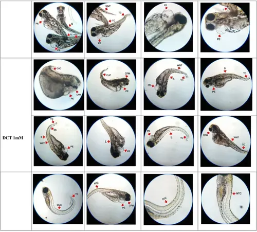
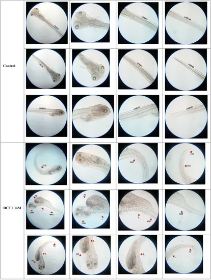
Representative images of developing embryos at 120 hpf in groups exposed to different concentrations of DCT. Figure illustrates the developmental process of embryos at 120 hpf in groups exposed to DCT ranging from concentration 250 µM to 1mM (n=20). In comparison to the control group, the embryos exposed to DCT showed multiple developmental defects in both cranial and caudal portion of neural tube.The deformities in developing neural tube are indicated by arrow marks. The deformities specifiedin group exposed to DCT 250 µM were kyphosis (K), spinal split (SS) and wavy neural tube (WNT). The deformities specified in group exposed to DCT 500 µM were pericardial edema (PE), lordosis (L), curly-up tail curvature (CUC), scoliosis (S), tail curvature (TC), craniofacial malformation (CFM), wavy neural tube (WNT) and neural tube curvature (NTC). The deformities specified in group exposed to DCT 750 µM were tail curvature (TC), pericardial edema (PE), neural tube curvature (NTC), curly-up tail curvature (CUC), pericardial edema (PE), spinal split (SS), curly-up tail curvature (CUC), lordosis (L), scoliosis (S) and wavy neural tube (WNT). The deformities specified in group exposed to DCT 1mM were curly-up tail curvature (CUC), pericardial edema (PE), craniofacial malformation (CFM), pericardial edema (PE), lordosis (L), tail curvature (TC), wavy neural tube (WNT), scoliosis (S), spinal split (SS) and neural tube curvature (NTC) PHENOTYPE:
|

The stacked column chart illustrates the percentage of developing embryos with neural tube defects at 120 hpf on exposure to different concentrations of decitabine (DCT). The percentage of phenotypic outcomes with neural tube defects in groups exposed to DCT 250 µM, 500 µM, 750 µM, and 1 mM was 10%, 65%, 85% and 100% respectively. A concentration dependent increase in percentage of phenotypic outcomes with neural tube defects was noted. Values are significantly different from control at 120 hpf. ns-non significant, ***P < 0.0001 (one-way ANOVA followed by Dunnett’s test), n=20 |
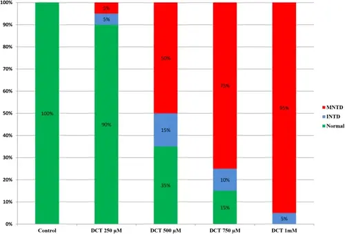
The stacked column chart illustrates the percentage of developing embryos with isolated neural tube defect (INTD) and multiple neural tube defects at 120 hpf in embryos exposed to different concentrations of decitabine (DCT) (n=20). The phenotypic outcomes with isolated neural tube defects (INTDs) noted in groups exposed to decitabine 250 µM, 500 µM, 750 µM and 1 mM was 5%, 15%, 10% and 5% respectively. The phenotypic outcomes with multiple neural tube defects (MNTDs) in groups exposed to decitabine 250 µM, 500 µM, 750 µM and 1mM was 5%, 50%, 75% and 95% respectively. A concentration dependent increase in severity of neural tube defect was noted in groups exposed to decitabine. With 95% of developing embryos suffering from multiple neural tube defects and 5% from isolated neural tube defects, all of the embryos in the group exposed to 1 mM decitabine had neural tube defects |
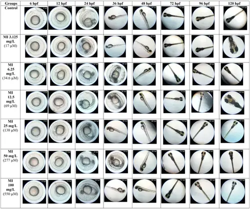
Embryonic development of groups exposed to different concentrations of MI. Figure illustrates the representative images of the developmental process of embryos in groups exposed to graded concentrations of MI ranging from 3.125 mg/L to 100 mg/L at various time intervals-hours post fertilization (hpf). The embryos exposed to different MI concentrations (3.125 mg/L, 6.25 mg/L, 12.5 mg/L, 25 mg/L, 50 mg/L, and 100 mg/L) showed normal development process, comparable to the control group |
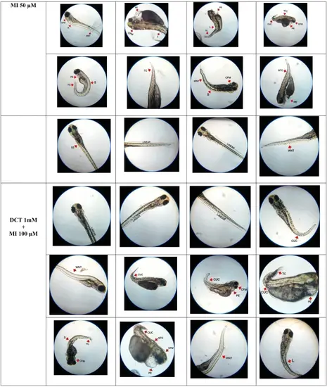
Representative images of developing embryos at 120 hpf in groups exposed to DCT alone and DCT with MI. At 120 hpf, all developing embryos in the DCT group showed abnormalities in the developing neural tube at multiple sites. The deformities specified were kyphosis (K), scoliosis (S), wavy neural tube (WNT), pericardial edema (PE), neural tube curvature (NTC), tail curvature (TC), craniofacial malformation (CFM), spinal split (SS) and curly-uptail curvature (CUC).Embryos in the DCT and MI 50 µM and 100 µM groups hadboth normal neural tube development and neural tube deformitiesat multiple sites.The deformities specified in embryos exposed to DCT and MI 50 µM were pericardial edema (PE), lordosis (L), neural tube curvature (NTC), kyphosis (K), craniofacial malformation (CFM), wavy neural tube (WNT), tail curvature (TC), curly-up tail curvature (CUC) and scoliosis (S). The deformities specified in embryos exposed to DCT and MI 100 µM were curly-up tail curvature (CUC), wavy neural tube (WNT), pericardial edema (PE), craniofacial malformation (CFM), tail curvature (TC), scoliosis (S), neural tube curvature (NTC), wavy neural tube (WNT) and lordosis (L). The deformities in developing neural tube are indicated by arrow marks |

The stacked column chart illustrates the percentage of developing embryos with neural tube defects at 120 hpf in groups exposed to decitabine alone and in the presence of myo-inositol (MI). The percentage of phenotypic outcomes with neural tube defects in group exposed to DCT 1 mM was 100% and in groups exposed to DCT 1 mM + MI 50 µM and DCT 1 mM + MI 100 µM, the percentage of phenotypic outcomes with neural tube defects was found to be 65% and 70% respectively. aValues are significantly different from control at 120 hpf, bValues are significantly different from DCT 1 mM group at 120 hpf. *P<0.05, **P<0.001,***P < 0.0001 (one-way ANOVA followed by Dunnett’s test), n=20 PHENOTYPE:
|
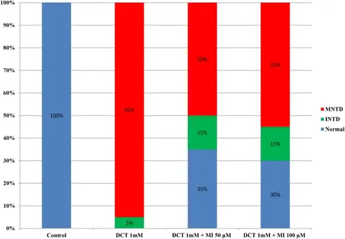
The stacked column chart illustrates the percentage of developing embryos with isolated and multiple neural tube defects at 120 hpf in groups exposed to decitabine alone and in presence of myo-inositol (n=20). The percentage of phenotypic outcomes with isolated neural tube defect in group exposed to DCT 1 mM was 5% and multiple neural tube defect was 95% with 100% incidence of neural tube defects, whereas in group exposed to DCT 1 mM + MI 50 µM, the percentage of phenotypic outcomes with isolated neural tube defect was 15% and the percentage of phenotypic outcomes with multiple neural tube defect was 50%. The remaining 35% were found normal. In group exposed to DCT 1 mM + MI 100 µM, the percentage of phenotypic outcomes with isolated neural tube defect was 15%, and the percentage of phenotypic outcomes with multiple neural tube defect was 55% and the remaining 30% appeared normal |
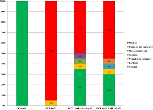
The stacked column chart illustrates the percentage of developing embryos with isolated neural tube defect (INTD) at 120 hpf in groups exposed to decitabine alone and in the presence of myo-inositol (n=20). The phenotypic outcomes with isolated neural tube defects (INTDs) noted in groups exposed decitabine 1 mM was lordosis (5%) and the remaining 95% suffered from multiple neural tube defects. In group exposed to dectabine 1 mM and myo-inositol 50 µM, the phenotypic outcomes with isolated neural tube defects were lordosis (5%), scoliosis (5%) and neural tube curvature (5%). 50% of developing embryos suffered from multiple neural tube defects and 35% was observed normal. In group exposed to dectabine 1 mM and myo-inositol 100 µM, the phenotypic outcomes with isolated neural tube defects were lordosis (5%), wavy neural tube (5%), curly up-tail curvature (5%). 55% of developing embryos suffered from multiple neural tube defects and 30% of developing embryos were observed normal |
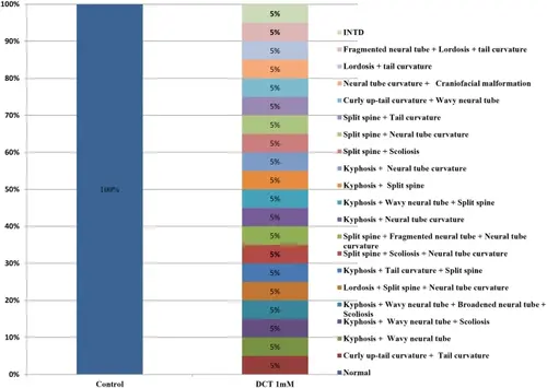
The stacked column chart illustrates the percentage of developing embryos with multiple deformities in neural tube (multiple neural tube defects) and isolated neural tube defect at 120 hpf in groups exposed to decitabine 1mM (n=20). The multiple neural tube defects noted were curly up-tail curvature + tail curvature (5%), kyphosis + wavy neural tube (5%), kyphosis + wavy neural tube + scoliosis (5%), kyposis + wavy neural tube + broadened neural tube + scoliosis (5%), lordosis + split spine + neural tube curvature (5%), kyphosis + tail curvature + split spine (5%), split spine + scoliosis + neural tube curvature (5%), split spine + fragmented neural tube + neural tube curvature (5%), kyphosis + neural tube curvature (5%), kyphosis + wavy neural tube + split spine (5%), kyphosis + split spine (5%), kyphosis + neural tube curvature (5%), split spine + scoliosis (5%), split spine + neural tube curvature (5%), split spine + tail curvature (5%), curly up-tail curvature + wavy neural tube (5%), neural tube curvature + craniofacial malformation (5%), lordosis + tail curvature (5%), fragmented neural tube + lordosis + tail curvature (5%). The remaining 5% of developing embryos suffered from isolated neural tube curvature (INTD) |
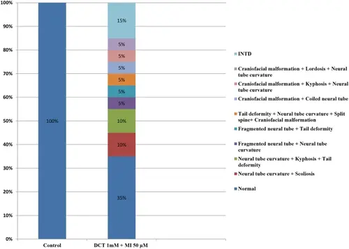
The stacked column chart illustrates the percentage of developing embryos with multiple deformities in neural tube (multiple neural tube defects) and isolated neural tube defect at 120 hpf in groups exposed to decitabine 1mM and myo-inositol 50µM (n=20). The multiple neural tube defects noted were neural tube curvature + scoliosis (10%), neural tube curvature + kyphosis + tail deformity (10%), fragmented neural tube + neural tube curvature (5%), fragmented neural tube + tail deformity (5%), tail deformity + neural tube curvature + split spine + craniofacial malformation (5%), craniofacial malformation + coiled neural tube (5%), craniofacial malformation + kyphosis + neural tube curvature (5%) and craniofacial malformation + lordosis + neural tube curvature (5%). The remaining 15% of developing embryos suffered from isolated neural tube defects (INTD) and 35% of developing embryos were observed normal |
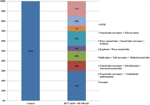
The stacked column chart illustrates the percentage of developing embryos with multiple deformities in neural tube (multiple neural tube defects) and isolated neural tube defect at 120 hpf in groups exposed to decitabine 1mM and myo-inositol 100µM (n=20). The multiple neural tube defects noted were neural tube curvature + craniofacial malformation (10%), neural tube curvature + tail deformity + narrowed neural tube (10%), split spine + tail curvature + kinked neural tube (5%), kyphosis + wavy neural tube (15%), wavy neural tube + neural tube curvature + scoliosis (5%) and neural tube curvature + tail curvature (10%). The remaining 15% of developing embryos suffered from isolated neural tube defects (INTD) and 30% of developing embryos were observed normal |
Representative images of alizarin red stained zebrafish larvae at 120 hpf exposed to DCT alone and DCT with MI (n=20). The figure illustrates the linearity of neural tube in control group larvae. The image of stained larvae in group exposed to decitabine 1mM show multiple deformities in neural tube. The deformities specified in images were curly up-tail curvature (CUC), tail curvature (TC), craniofacial malformation (CFM), pericardial edema (PE), lordosis (L) and wavy neural tube (WNT). In group exposed to DCT 1 mM and MI 50 µM, the neural tube of 35% of the total number of larvae exposed were found linear without any deformities as shown in figure. In group exposed to DCT 1 mM and MI 100 µM, the neural tube of 30% of the total number of larvae exposed were found linear without any deformities as shown in figure. The deformity specified in image in group exposed to DCT 1 mM and MI 100 µM is spinal split (SS) |
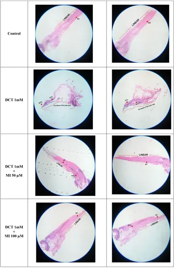
Representative histopathological images (10X) of paraformaldehyde fixed zebrafish larvae at 120 hpf which are exposed to decitabine (DCT) alone and in presence of myo-inositol (MI) (n=20). Histology of control group larvae shows neural tube (NT) with normal architecture. The histology of decitabine (DCT) 1 mM exposed larvae show curvature in neural tube (NTC) with curved body axis and abnormal morphology. The group exposed to DCT 1 mM and MI 50 µM show linear caudal region with a mild curvature in cranial region. The group exposed to DCT 1 mM and MI 100 µM show linear caudal region with a mild curvature in neural tube. The red arrow marks indicates the neural tube and neural tube curvatures (NTC). The curvature in body axis is indicated in group exposed to decitabine as a curved red line. Linearity in body axis is indicated in control group and group exposed to decitabine and myo-inositol is indicated as red straight line |