
FIGURE
Fig. 4
- ID
- ZDB-FIG-250421-46
- Publication
- Rajesh et al., 2025 - Protective Effect of myo-Inositol Against Decitabine-Induced Neural Tube Defects in Embryonic Zebrafish
- Other Figures
- All Figure Page
- Back to All Figure Page
Fig. 4
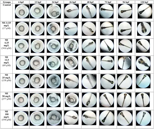
Embryonic development of groups exposed to different concentrations of MI. Figure illustrates the representative images of the developmental process of embryos in groups exposed to graded concentrations of MI ranging from 3.125 mg/L to 100 mg/L at various time intervals-hours post fertilization (hpf). The embryos exposed to different MI concentrations (3.125 mg/L, 6.25 mg/L, 12.5 mg/L, 25 mg/L, 50 mg/L, and 100 mg/L) showed normal development process, comparable to the control group |
Expression Data
Expression Detail
Antibody Labeling
Phenotype Data
| Fish: | |
|---|---|
| Condition: | |
| Observed In: | |
| Stage Range: | Shield to Day 5 |
Phenotype Detail
Acknowledgments
This image is the copyrighted work of the attributed author or publisher, and
ZFIN has permission only to display this image to its users.
Additional permissions should be obtained from the applicable author or publisher of the image.
Full text @ Neurotox Res