
Fig. 13
- ID
- ZDB-FIG-250421-55
- Publication
- Rajesh et al., 2025 - Protective Effect of myo-Inositol Against Decitabine-Induced Neural Tube Defects in Embryonic Zebrafish
- Other Figures
- All Figure Page
- Back to All Figure Page
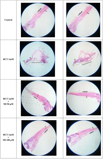
Representative histopathological images (10X) of paraformaldehyde fixed zebrafish larvae at 120 hpf which are exposed to decitabine (DCT) alone and in presence of myo-inositol (MI) (n=20). Histology of control group larvae shows neural tube (NT) with normal architecture. The histology of decitabine (DCT) 1 mM exposed larvae show curvature in neural tube (NTC) with curved body axis and abnormal morphology. The group exposed to DCT 1 mM and MI 50 µM show linear caudal region with a mild curvature in cranial region. The group exposed to DCT 1 mM and MI 100 µM show linear caudal region with a mild curvature in neural tube. The red arrow marks indicates the neural tube and neural tube curvatures (NTC). The curvature in body axis is indicated in group exposed to decitabine as a curved red line. Linearity in body axis is indicated in control group and group exposed to decitabine and myo-inositol is indicated as red straight line |