
Fig. 12
- ID
- ZDB-FIG-250421-54
- Publication
- Rajesh et al., 2025 - Protective Effect of myo-Inositol Against Decitabine-Induced Neural Tube Defects in Embryonic Zebrafish
- Other Figures
- All Figure Page
- Back to All Figure Page
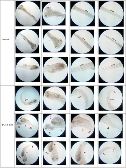
Representative images of alizarin red stained zebrafish larvae at 120 hpf exposed to DCT alone and DCT with MI (n=20). The figure illustrates the linearity of neural tube in control group larvae. The image of stained larvae in group exposed to decitabine 1mM show multiple deformities in neural tube. The deformities specified in images were curly up-tail curvature (CUC), tail curvature (TC), craniofacial malformation (CFM), pericardial edema (PE), lordosis (L) and wavy neural tube (WNT). In group exposed to DCT 1 mM and MI 50 µM, the neural tube of 35% of the total number of larvae exposed were found linear without any deformities as shown in figure. In group exposed to DCT 1 mM and MI 100 µM, the neural tube of 30% of the total number of larvae exposed were found linear without any deformities as shown in figure. The deformity specified in image in group exposed to DCT 1 mM and MI 100 µM is spinal split (SS) |