FIGURE SUMMARY
Title

Effect of nonsense-mediated mRNA decay factor SMG9 deficiency on premature aging in zebrafish

Authors
Lai, S., Shiraishi, H., Sebastian, W.A., Shimizu, N., Umeda, R., Ikeuchi, M., Kiyota, K., Takeno, T., Miyazaki, S., Yano, S., Shimada, T., Yoshimura, A., Hanada, R., Hanada, T.
Source
Full text @ Commun Biol

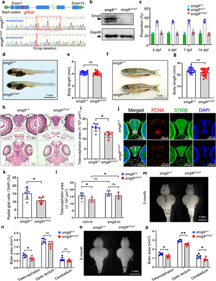
Smg9-deficient zebrafish display brain malformations.

a Genomic locus diagram depicting the zebrafish smg9 gene and its 10-nucleotide deletion using CRISPR/Cas9. The red box indicates the mutated region in smg9oi7/oi7 zebrafish. b Western blot analyses of lysates from smg9+/+ and smg9oi7/oi7 larvae at 2 dpf to determine the Smg9 protein levels. c The percentage of smg9+/+, smg9oi7/+, and smg9oi7/oi7 zebrafish were determined from 2 to 14 dpf. d Representative images of smg9+/+ and smg9oi7/oi7 larvae at 14 dpf. Scale bar: 1 mm. e Body length of smg9+/+ and smg9oi7/oi7 larvae at 14 dpf. f Representative images of smg9+/+ and smg9oi7/oi7 fish at 3 mpf. Scale bar: 1 cm. g Body length of smg9+/+ and smg9oi7/oi7 zebrafish at 3 mpf. h Representative images of H&E staining of transverse and coronal sections of smg9+/+ and smg9oi7/oi7 larvae at 14 dpf. Scale bar: 50 µm in the upper panel and 100 µm in the lower panel. i Telencephalon areas of smg9+/+ and smg9oi7/oi7 larvae at 14 dpf were measured using transverse sections of H&E images. j Representative images of immunostaining of 14 dpf larval brain tissue with PCNA (red), S100β (green), and DAPI (blue). Scale bar: 100 µm. k Graph shows PCNA/S100β double-positive radial glial cells among DAPI-positive cells in the telencephalon of smg9+/+ and smg9oi7/oi7 larvae at 14 dpf. l Telencephalon size of 14 dpf smg9+/+ and smg9oi7/oi7 larvae injected with control (con-in) or zebrafish smg9 mRNA (smg9-in). m Representative images of the dissected brains of smg9+/+ and smg9oi7/oi7 zebrafish at 3 mpf. Scale bar: 1 mm. n Quantification of the areas of the telencephalon, optic tectum, and cerebellum in smg9+/+ and smg9oi7/oi7 zebrafish at 3 mpf. o Representative images of dissected brains of smg9+/+ and smg9oi7/oi7 zebrafish at 6 mpf. Scale bar: 1 mm. p Quantification of the areas of the telencephalon, optic tectum, and cerebellum in smg9+/+ and smg9oi7/oi7 zebrafish at 6 mpf. Error bars indicate SD. *P < 0.05, **P < 0.01, and ***P < 0.001. ns: not significant.

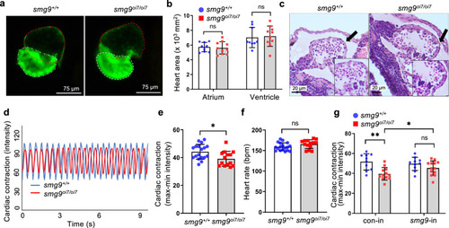
Impaired cardiac contractions in Smg9-deficient zebrafish.

a Representative image of the heart of cmlc2:EGFP zebrafish. The red and white dotted lines indicate the atrium and ventricle, respectively. b Quantification of the atrium and ventricle areas in smg9+/+ and smg9oi7/oi7 zebrafish at 6 dpf. c Representative images of hematoxylin and eosin staining of smg9+/+ and smg9oi7/oi7 hearts at 6 dpf. d Representative dynamic pixel change patterns of the heart in cmlc2:EGFP zebrafish, showing the heartbeat and cardiac contraction of smg9+/+ and smg9oi7/oi7 zebrafish. e Quantification of cardiac contractions in smg9+/+ and smg9oi7/oi7 zebrafish larvae at 6 dpf. f Quantification of the heart rates of smg9+/+ and smg9oi7/oi7 zebrafish larvae at 6 dpf. g Quantification of cardiac contractions of 6 dpf smg9+/+ and smg9oi7/oi7 larvae injected with control (con-in) or zebrafish smg9 mRNA (smg9-in). Error bars indicate SD. bpm: beats per minute. *P < 0.05, **P < 0.01, and ***P < 0.001. ns: not significant.

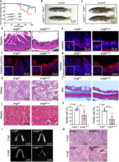
Premature aging phenotype in various tissues in Smg9-deficient zebrafish.

a Kaplan–Meier survival curve analysis showing the lifespan of smg9+/+ (n = 20), smg9oi7/+ (n = 20), and smg9oi7/oi7 (n = 20) zebrafish. b, c Representative images of gross appearance of smg9+/+ and smg9oi7/oi7 zebrafish at 3 mpf and 6 mpf. Scale bar: 5 mm. d Representative H&E images of the intestine showing tissue morphology in smg9+/+ and smg9oi7/oi7 zebrafish at 6 mpf. Scale bar: 50 μm. eg Representative images of immunofluorescence staining for PCNA (e), γH2AX (f), and cleaved caspase-3 (g) in the intestinal segments of smg9+/+ and smg9oi7/oi7 zebrafish at 6 mpf. Scale bar: 50 μm. h Representative H&E images of the kidney tissue morphology in smg9+/+ and smg9oi7/oi7 zebrafish at 6 mpf. Scale bar: 50 μm. i, j Representative images of Masson’s trichrome staining of smg9+/+ and smg9oi7/oi7 skin (i) and muscle (j) at 6 mpf. Scale bar: 10 μm (i). Scale bar: 20 μm (j). k Fertility rate of smg9+/+ and smg9oi7/oi7 zebrafish at 4 mpf and 7 mpf, as determined by mating smg9+/+ or smg9oi7/oi7 male zebrafish with smg9+/+ female zebrafish and counting embryo survival after 12 hpf. l Representative images of the morphological features of testes in smg9+/+ and smg9oi7/oi7 zebrafish at 3 mpf and 6 mpf. Scale bar: 2.5 mm. m Representative image of H&E staining of testes tissue in smg9+/+ and smg9oi7/oi7 at 3 mpf and 6 mpf. Sperm (black arrow) was absent in the 6 mpf smg9oi7/oi7 zebrafish. Scale bar: 50 μm. Error bars indicate SD. *P < 0.05, ** P < 0.01, and *** P < 0.001. ns: not significant.

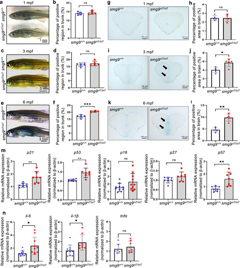
Age-related staining and markers in Smg9-deficient zebrafish.

a, b SA-β-gal staining of the skin of smg9+/+ and smg9oi7/oi7 zebrafish at 1 mpf. Scale bar: 1 mm (a). Quantification of the percentage of positive areas in the trunk at 1 mpf (b). c, d SA-β-gal staining of the skin of smg9+/+ and smg9oi7/oi7 zebrafish at 3 mpf (c). Quantification of the percentage of positive areas in the trunk at 3 mpf. Scale bar: 3 mm (d). e, f SA-β-gal staining of the skin of smg9+/+ and smg9oi7/oi7 zebrafish at 6 mpf. Scale bar: 3 mm (e). Quantification of the percentage of positive areas in the trunk at 6 mpf (f). g, h SA-β-gal staining of the frozen brain sections of smg9+/+ and smg9oi7/oi7 zebrafish at 1 mpf. Scale bar: 50 μm (g). Quantification of the percentage of positive areas in the brain at 1 mpf (h). i, j SA-β-gal staining of the frozen brain sections of smg9+/+ and smg9oi7/oi7 zebrafish at 3 mpf. The arrows indicate positive regions. Scale bar: 100 μm (i). Quantification of the percentage of positive areas in the brain at 3 mpf (j). k, l SA-β-gal staining of the frozen brain sections of smg9+/+ and smg9oi7/oi7 zebrafish at 6 mpf. The arrows indicate positive regions. Scale bar: 100 μm (k). Quantification of the percentage of positive areas in the brain at 6 mpf (l). m Quantitative PCR analysis of p21, p53, p16, p27, and p57 expression in smg9+/+ and smg9oi7/oi7 zebrafish larvae at 14 dpf. n Quantitative PCR analysis of il-6, il-1β and tnfα expression in smg9+/+ and smg9oi7/oi7 zebrafish larvae at 14 dpf. Error bars indicate SD. *P < 0.05, ** P < 0.01, and *** P < 0.001. ns not significant.

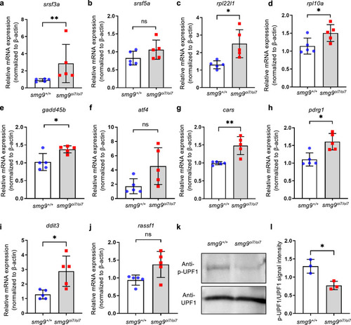
Upregulation of endogenous targets of nonsense-mediated mRNA decay (NMD) in Smg9-deficient zebrafish.

aj Quantitative PCR analysis of endogenous target genes of NMD in smg9+/+ and smg9oi7/oi7 larvae at 14 dpf. k Western blot analysis of lysates from smg9+/+ and smg9oi7/oi7 larvae at 2 dpf to determine the total FLAG-UPF1 (UPF1) and phosphorylated FLAG-UPF1(p-UPF1) levels. l Quantification of the total FLAG-UPF1 (UPF1) and phosphorylated FLAG-UPF1 (p-UPF1) levels in three independent experiments. Error bars indicate SD. *P < 0.05, ** P < 0.01, and *** P < 0.001. ns: not significant.

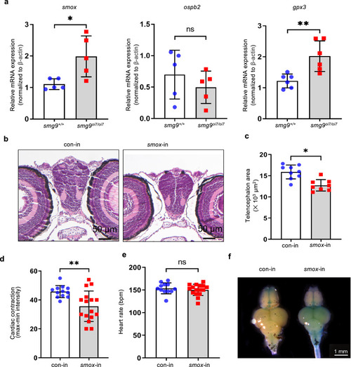
Smox overexpression is sufficient for brain and heart defects in Smg9-deficient zebrafish.

a Quantitative PCR analysis of endogenous target genes of nonsense-mediated mRNA decay in smg9+/+ and smg9oi7/oi7 larvae at 14 dpf. bfSmox mRNA was injected into single-cell-stage fertilized eggs derived from the smg9+/+ zebrafish. The telencephalon area at 14 dpf and the heart function at 6 dpf in zebrafish larvae were evaluated. b Representative images of H&E staining of smox mRNA-injected (smox-in) and control-injected (con-in) larvae at 14 dpf. Scale bar: 50 μm. c Quantification of the telencephalon area of smox-in and con-in larvae at 14 dpf was performed using hematoxylin and eosin staining. d Quantification of cardiac contractions in smox-in and con-in larvae at 6 dpf. e Quantification of the heart rate of smox-in and con-in larvae at 6 dpf. f SA-β-gal staining of the brain of smox-in and con-in zebrafish at 4 mpf. Error bars indicate SD. bpm: beats per minute. *P < 0.05, ** P < 0.01, and *** P < 0.001. ns: not significant.

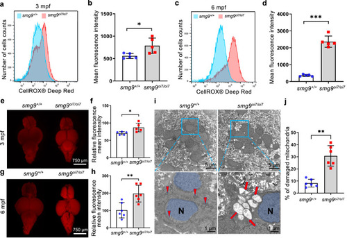
ROS and acrolein accumulation in Smg9-deficient zebrafish.

a, c Representative flow cytometry histograms of CellROX fluorescence in smg9+/+ and smg9oi7/oi7 zebrafish at 3 mpf (a) and 6 mpf (c). b, d Mean fluorescence intensity of CellROX Deep Red in the brains of smg9+/+ and smg9oi7/oi7 zebrafish at 3 mpf (b) and 6 mpf (d). e, g Representative images of AcroleinRED staining of smg9+/+ and smg9oi7/oi7 zebrafish brains at 3 mpf (e) and 6 mpf (g). f, h Relative mean fluorescence intensities of smg9+/+ and smg9oi7/oi7 zebrafish brains at 3 mpf (f) and 6 mpf (h). i, j Representative images of transmission electron microscopy of smg9+/+ and smg9oi7/oi7 zebrafish brains at 6 mpf. The red arrowhead and red arrow indicate normal and damaged mitochondria, respectively. N Nucleus. Scale bar: 5 µm in the upper panel and 1 µm in the lower panel (i). The density of damaged mitochondria within a defined area (0.25 mm2) in the telencephalon region of six individual zebrafish brains (j). Error bars indicate SD. *P < 0.05, ** P < 0.01, and *** P < 0.001. ns: not significant.

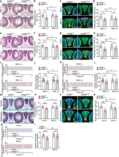
Smox is required for brain and heart defects in Smg9-deficient zebrafish.

a Representative image of hematoxylin and eosin staining of smg9+/+ and smg9oi7/oi7 larvae after treatment with or without MDL72527. Scale bar: 50 µm. b Quantification of the telencephalon area of smg9+/+ and smg9oi7/oi7 larvae after treatment with or without MDL72527. c Representative images of immunostaining for PCNA (red), S100β (green), and DAPI (blue) for smg9+/+ and smg9oi7/oi7 larvae after treatment with or without MDL72527. Scale bar: 50 µm. d Quantification of PCNA/S100β double-positive radial glial cells among DAPI-positive cells in the telencephalon of smg9+/+ and smg9oi7/oi7 larvae after treatment with or without MDL72527. e Representative images of hematoxylin and eosin staining of smg9+/+ and smg9oi7/oi7 larvae after treatment with or without N-acetylcysteine (NAC). Scale bar: 50 µm. f Quantification of the telencephalon area of smg9+/+ and smg9oi7/oi7 larvae after treatment with or without NAC. Scale bar: 50 µm. g Representative images of immunostaining for PCNA (red), S100β (green), and DAPI (blue) for smg9+/+ and smg9oi7/oi7 larvae after treatment with or without NAC. Scale bar: 50 µm. h Quantification of PCNA/S100β double-positive radial glial cells among DAPI-positive cells in the telencephalon of smg9+/+ and smg9oi7/oi7 larvae, after treatment with or without NAC. i Representative dynamic pixel change patterns of the heart showing cardiac contractions of smg9+/+ and smg9oi7/oi7 zebrafish after treatment with or without MDL72527. j Quantification of cardiac contractions in smg9+/+ and smg9oi7/oi7 zebrafish larvae after treatment with or without MDL72527. k Representative dynamic pixel change patterns of the heart showing cardiac contractions of smg9+/+ and smg9oi7/oi7 zebrafish after treatment with or without NAC. l Quantification of cardiac contractions in smg9+/+ and smg9oi7/oi7 zebrafish larvae after treatment with or without NAC. m Representative images of hematoxylin and eosin staining of smg9+/+ and smg9oi7/oi7 larvae injected with control (con-in) or smox morpholino (MO-in). Scale bar: 50 µm. n Quantification of the telencephalon area of smg9+/+ and smg9oi7/oi7 larvae injected with control (con-in) or smox morpholinos (MO-in). o Representative images of immunostaining with PCNA (red), S100β (green), and DAPI (blue) for smg9+/+ and smg9oi7/oi7 larvae injected with control (con-in) or smox morpholino (MO-in). Scale bar: 50 µm. p Quantification of PCNA/S100β double-positive radial glial cells among DAPI-positive cells in the telencephalon of smg9+/+ and smg9oi7/oi7 larvae injected with control (con-in) or smox morpholino (MO-in). q Representative dynamic pixel change patterns of the heart showing cardiac contraction of smg9+/+ and smg9oi7/oi7 zebrafish injected with control (con-in) or smox morpholino (MO-in). r Quantification of cardiac contractions in smg9+/+ and smg9oi7/oi7 zebrafish larvae injected with control (con-in) or smox morpholino (MO-in). Error bars indicate SD. *P < 0.05, ** P < 0.01, and *** P < 0.001. ns: not significant.

Acknowledgments
This image is the copyrighted work of the attributed author or publisher, and ZFIN has permission only to display this image to its users. Additional permissions should be obtained from the applicable author or publisher of the image. Full text @ Commun Biol