IMAGE

Fig. 1

ID
ZDB-IMAGE-240531-136
Source
Figures for Lai et al., 2024
Image
Figure Caption

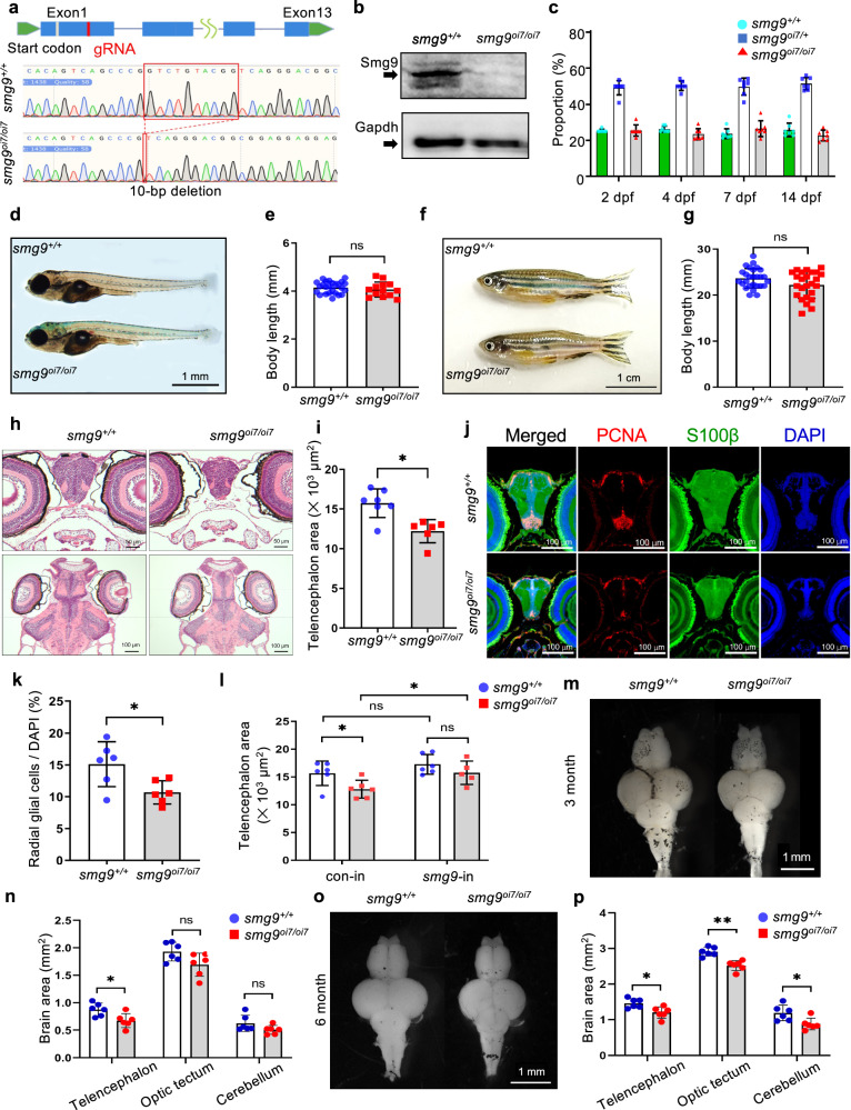
Fig. 1 Smg9-deficient zebrafish display brain malformations.

a Genomic locus diagram depicting the zebrafish smg9 gene and its 10-nucleotide deletion using CRISPR/Cas9. The red box indicates the mutated region in smg9oi7/oi7 zebrafish. b Western blot analyses of lysates from smg9+/+ and smg9oi7/oi7 larvae at 2 dpf to determine the Smg9 protein levels. c The percentage of smg9+/+, smg9oi7/+, and smg9oi7/oi7 zebrafish were determined from 2 to 14 dpf. d Representative images of smg9+/+ and smg9oi7/oi7 larvae at 14 dpf. Scale bar: 1 mm. e Body length of smg9+/+ and smg9oi7/oi7 larvae at 14 dpf. f Representative images of smg9+/+ and smg9oi7/oi7 fish at 3 mpf. Scale bar: 1 cm. g Body length of smg9+/+ and smg9oi7/oi7 zebrafish at 3 mpf. h Representative images of H&E staining of transverse and coronal sections of smg9+/+ and smg9oi7/oi7 larvae at 14 dpf. Scale bar: 50 µm in the upper panel and 100 µm in the lower panel. i Telencephalon areas of smg9+/+ and smg9oi7/oi7 larvae at 14 dpf were measured using transverse sections of H&E images. j Representative images of immunostaining of 14 dpf larval brain tissue with PCNA (red), S100β (green), and DAPI (blue). Scale bar: 100 µm. k Graph shows PCNA/S100β double-positive radial glial cells among DAPI-positive cells in the telencephalon of smg9+/+ and smg9oi7/oi7 larvae at 14 dpf. l Telencephalon size of 14 dpf smg9+/+ and smg9oi7/oi7 larvae injected with control (con-in) or zebrafish smg9 mRNA (smg9-in). m Representative images of the dissected brains of smg9+/+ and smg9oi7/oi7 zebrafish at 3 mpf. Scale bar: 1 mm. n Quantification of the areas of the telencephalon, optic tectum, and cerebellum in smg9+/+ and smg9oi7/oi7 zebrafish at 3 mpf. o Representative images of dissected brains of smg9+/+ and smg9oi7/oi7 zebrafish at 6 mpf. Scale bar: 1 mm. p Quantification of the areas of the telencephalon, optic tectum, and cerebellum in smg9+/+ and smg9oi7/oi7 zebrafish at 6 mpf. Error bars indicate SD. *P < 0.05, **P < 0.01, and ***P < 0.001. ns: not significant.

Acknowledgments
This image is the copyrighted work of the attributed author or publisher, and ZFIN has permission only to display this image to its users. Additional permissions should be obtained from the applicable author or publisher of the image. Full text @ Commun Biol