
- Title
-
Rab8, Rab11, and Rab35 coordinate lumen and cilia formation during zebrafish left-right organizer development
- Authors
- Aljiboury, A.A., Ingram, E., Krishnan, N., Ononiwu, F., Pal, D., Manikas, J., Taveras, C., Hall, N.A., Da Silva, J., Freshour, J., Hehnly, H.
- Source
- Full text @ PLoS Genet.
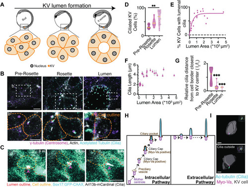
(A) Model depicting KV lumen formation across developmental stages of the zebrafish embryo. (B) Confocal micrographs of KV developmental stages with cilia (acetylated-tubulin, cyan), centrosome (γ-tubulin, magenta), and actin (phalloidin, gray). Scale bar, 10 μm. (b’) Magnified insets from (B) depicting centrosome and cilia positioning in KV cells at different KV developmental stages. Scale bar, 7 μm. (C) KV cell building and extending a cilium (Arl13b-mCardinal, gray) into the lumen of the KV. KV plasma membranes (Sox17:GFP-CAAX, cyan) shown. Scale bar, 5 μm. (D) Percentage of ciliated KV cells at the different KV developmental stages shown as a violin plot with median (yellow line). One way ANOVA across KV developmental stages, n>7 embryos, **p<0.01. (E) Scatter plot demonstrating the percentage of KV cells with lumenal cilia per embryo in relation to KV lumen area. n = 29 embryos. Goodness of fit R2 = 0.8577. (F) Scatter plot depicting average cilia length within KV cells per embryo across n = 29 embryos in relation to lumen area. Error bars, ± SEM. |
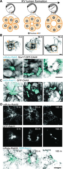
(A) Model depicting rosette, early lumen and late lumen stages of KV development being examined in live embryos represented in panels (B-D). (B-D) Representative images from live confocal videos of ectopically expressed mRuby-Rab8 (cyan, B), mCherry-Rab11 (cyan, C), endogenously tagged GFP-Rab11 (gray, cyan in merge, D) and ectopically expressed mRuby-Rab35 (gray, inverted gray in merge, D) localization in KV cells marked by GFP-CAAX (inverted gray, B) during KV lumen formation. Scale bar, 10μm. Refer to S2 Video. |
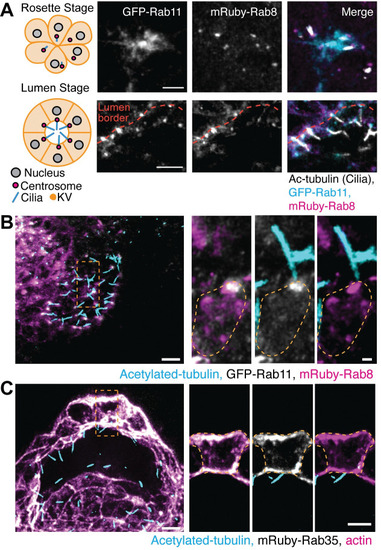
(A) Left, model of KV developmental stages, rosette (top) and lumen (bottom), with centrosome (magenta) and cilia (cyan) positioning. Right, confocal micrographs with GFP-Rab11 (gray, cyan in merge), mRuby-Rab8 (gray, magenta in merge), and cilia (acetylated-tubulin, gray), shown. Scale bar, 10 μm. (B-C) Confocal micrographs of KV lumen stage with cilia (acetylated-tubulin, cyan), GFP-Rab11 (gray, B), mRuby-Rab8 (magenta, B), mRuby-Rab35 (gray, C), and actin (magenta, C). Scale bar, 2 μm. |
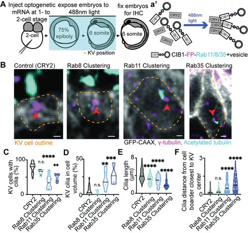
(A) A model depicting the use of optogenetics to acutely block Rab GTPase-associated trafficking events during KV developmental stages. (B) Confocal micrographs of cilia (acetylated tubulin, cyan) in CRY2 (control), Rab8-, Rab11-, and Rab35-clustered Sox17:GFP-CAAX embryos (gray). Centrosomes denoted by γ-tubulin (magenta). Clusters not shown. Yellow dashed lines are KV cell cortical membranes. Orange arrow, centrosome. Scale bar, 2μm. (C-F) Violin plots of percentage of KV cells with cilia (C), percentage of KV cilia in cell volume (D), cilia length (E), and the relative distance of cilia from the cell boarder closest to KV center (F). One way ANOVA with Dunnett’s multiple comparison to CRY2 (control) was performed. n>4 embryos, n.s. not significant, **p<0.01, ***p<0.001, ****p<0.0001. Statistical results detailed in S1 Table. |
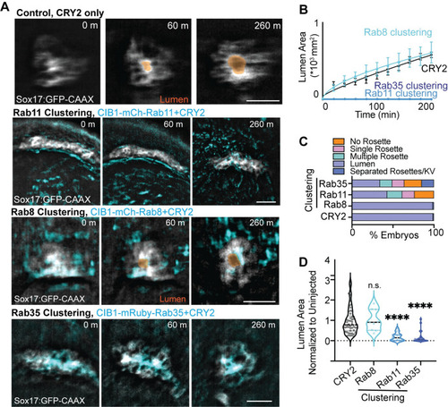
(A) Optogenetic clustering of Rab11 and Rab35 blocks KV lumen formation compared to CRY2 control and Rab8. Imaged on an automated fluorescent stereoscope. Scale bar, 50 μm. KV marked with Sox17:GFP-CAAX, lumens highlighted in orange, clusters shown in cyan. Refer to S3 Video. (B) KV lumen area over time (±SEM for n = 3 embryos per condition) in control (CRY2 injection) and Rab8, Rab11 and Rab35 clustering conditions. (C) KV morphologies characterized from optogenetically-clustered then fixed embryos at 12 SS (12 hpf). n>47 embryos per condition measured across n>9 clutches. (D) Violin plot depicting lumen area from Rab8, Rab11, and Rab35 clustering conditions normalized to uninjected control values. Dots represent individual KV values. Median denoted by line. One-way ANOVA with Dunnett’s multiple comparison test, compared to CRY2. n>9 embryos, n.s. not significant, ****p<0.0001. Statistical results detailed in S1 Table. |
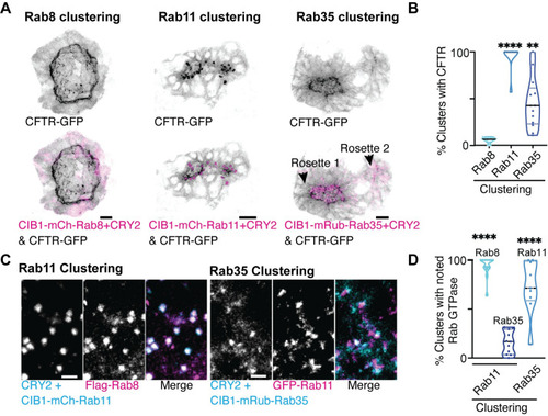
(A) Optogenetic clustering of Rab11, Rab8, and Rab35 (magenta) in KV cells. Localization with CFTR-GFP (inverted gray) is shown. Scale bar, 20 μm. (B) Violin plots depicting percent of optogenetic clusters that colocalize with CFTR. n>9 embryos, **p<0.01, ****p<0.0001. (C) Optogenetic clustering of CIB1-mCherry-Rab11 and CIB1-mRuby-Rab35 (cyan). CIB1-mCherry-Rab11 clusters localization with Flag-Rab8 (magenta) or CIB1-mRuby-Rab35 clusters with GFP-Rab11 (magenta) shown. Scale bar, 7 μm. (D) Violin plot depicting percent of optogenetic clusters that colocalize with Rab8, Rab35, or Rab11. n>9 embryos, ****p<0.0001. Statistical results detailed in S1 Table. |
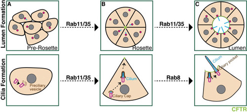
Centrosome depicted in magenta, cilia in cyan and CFTR in green. (A-B) At the pre-rosette stage (A) a proportion of centrosomes start to assemble cilia that then reposition towards the center of the KV at the rosette stage (B) in a Rab11 and Rab35 dependent manner. In (B), Rab11 and Rab35 mediate CFTR transport to the apical membrane. (C) The rosette stage then transitions to a lumen stage where most of the centrosomes locate at the CFTR-positive apical membrane and extend their cilia into the lumen where cilia elongate to their full length in a Rab8 dependent manner. |