
Image
|
Figure Caption
Fig 3
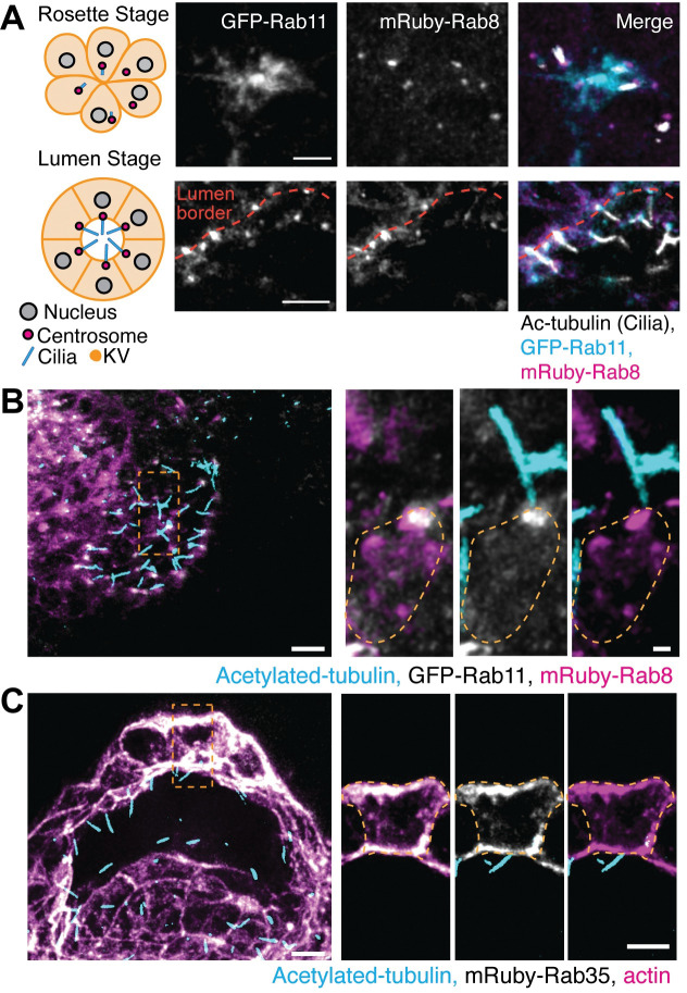
(A) Left, model of KV developmental stages, rosette (top) and lumen (bottom), with centrosome (magenta) and cilia (cyan) positioning. Right, confocal micrographs with GFP-Rab11 (gray, cyan in merge), mRuby-Rab8 (gray, magenta in merge), and cilia (acetylated-tubulin, gray), shown. Scale bar, 10 μm. (B-C) Confocal micrographs of KV lumen stage with cilia (acetylated-tubulin, cyan), GFP-Rab11 (gray, B), mRuby-Rab8 (magenta, B), mRuby-Rab35 (gray, C), and actin (magenta, C). Scale bar, 2 μm.
Acknowledgments
This image is the copyrighted work of the attributed author or publisher, and
ZFIN has permission only to display this image to its users.
Additional permissions should be obtained from the applicable author or publisher of the image.
Full text @ PLoS Genet.