
Fig 1
- ID
- ZDB-FIG-230526-55
- Publication
- Aljiboury et al., 2023 - Rab8, Rab11, and Rab35 coordinate lumen and cilia formation during zebrafish left-right organizer development
- Other Figures
- All Figure Page
- Back to All Figure Page
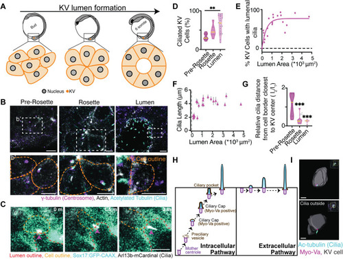
(A) Model depicting KV lumen formation across developmental stages of the zebrafish embryo. (B) Confocal micrographs of KV developmental stages with cilia (acetylated-tubulin, cyan), centrosome (γ-tubulin, magenta), and actin (phalloidin, gray). Scale bar, 10 μm. (b’) Magnified insets from (B) depicting centrosome and cilia positioning in KV cells at different KV developmental stages. Scale bar, 7 μm. (C) KV cell building and extending a cilium (Arl13b-mCardinal, gray) into the lumen of the KV. KV plasma membranes (Sox17:GFP-CAAX, cyan) shown. Scale bar, 5 μm. (D) Percentage of ciliated KV cells at the different KV developmental stages shown as a violin plot with median (yellow line). One way ANOVA across KV developmental stages, n>7 embryos, **p<0.01. (E) Scatter plot demonstrating the percentage of KV cells with lumenal cilia per embryo in relation to KV lumen area. n = 29 embryos. Goodness of fit R2 = 0.8577. (F) Scatter plot depicting average cilia length within KV cells per embryo across n = 29 embryos in relation to lumen area. Error bars, ± SEM. |